





Subvisible particles, those 2–100 µm in diameter, in protein, cell, and gene therapies and other parenteral drug products can pose risks to product safety, quality, and efficacy. To help mitigate these risks, the United States Pharmacopeia (USP) requires manufacturers to monitor the subvisible particle content of biotherapeutics and other parenteral drug products. These requirements are found in USP <787>, <788>, and <789>.
USP also provides recommendations for subvisible particle measurements in USP <1787>1 and <1788>2. Because these chapter numbers are greater than 1000, they are informational chapters and do not contain mandatory tests. However, following their recommendations can help scientists more effectively detect, characterize, and control subvisible particle populations.
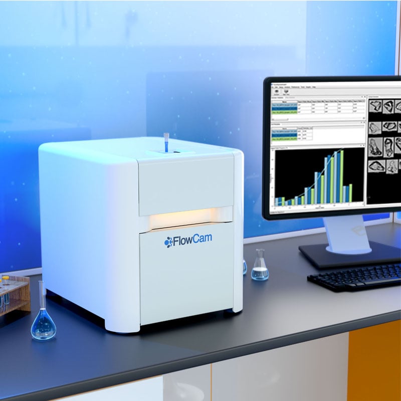
This article provides an overview of USP <1787> and <1788> and the particle-monitoring strategies they recommend. It also discusses how FlowCam, a flow imaging microscopy (FIM) instrument, can provide particle images and morphological measurements that complement compendial particle-counting methods.
Download the White Paper: USP Reference Standards for Subvisible Particulate Matter
USP <1787>: Measurement of Subvisible Particulate Matter in Therapeutic Protein Injections
USP <1787> focuses on strategies for characterizing subvisible particles in therapeutic protein products. Protein drug substances can form aggregate particles. These are considered an inherent particle type because they arise from the nature of the drug substance and may be an expected attribute of the product. In contrast, intrinsic particles originate from the product, its container, or the manufacturing process, while extrinsic particles originate outside the normal manufacturing process.
Protein aggregates present particular analytical challenges. Their low refractive index can make them difficult to detect using methods that depend strongly on optical contrast. Their flexible, fragile structures may also be altered or lost during filtration and other sample-preparation steps.
USP <1787> recommends using multiple complementary and orthogonal analytical techniques in addition to the compendial light obscuration and microscopic particle count methods. Techniques discussed in the chapter include flow imaging microscopy, electrical sensing zone, and electron microscopy. The chapter also recommends evaluating particles in the 1–10 µm range. Therapeutic protein products may contain substantial particle populations below the ≥10 µm reporting threshold used in compendial testing. USP <1787> notes that these particles are important because protein aggregates in this size range may contribute to an unwanted immune response, and changes in particle concentration can serve as an early indicator of product stability issues. Because counts in this size range can be highly variable, meaningful trend analysis requires appropriate controls, adequate sampling, and good analytical practice.
USP <1787> also discusses the importance of distinguishing protein aggregates from other particle types, including silicone oil droplets. Particle images and morphological measurements can provide information that particle counts alone cannot.
Although USP <1787> primarily applies to therapeutic protein injections, principles described in the chapter may also support particle characterization in other parenteral products. For example, imaging techniques may be used to examine viral or nonviral vector aggregates in gene therapies, as well as cell clusters and debris in cell-based products.
Why Does USP <1787> Recommend Orthogonal Particle Analysis?
No single particle-analysis method provides a complete description of every particle population. Individual techniques detect particles according to different physical properties, such as light blockage, electrical resistance, optical contrast, or morphology. They may therefore produce different results when used to analyze the same sample.
An orthogonal analytical strategy uses methods based on different measurement principles. Comparing information from multiple methods can help scientists evaluate particle concentration and size while also investigating properties such as shape, transparency, structure, and possible origin.
This approach is particularly valuable for therapeutic proteins because protein aggregates may be difficult to distinguish from silicone oil droplets, fibers, glass, or other intrinsic and extrinsic particles using particle counts alone.
Learn more about the role of complementary methods in our article on orthogonal methods for particle analysis.
USP <1788>: Methods for the Determination of Subvisible Particulate Matter
USP <1788> provides recommendations for implementing methods used to measure subvisible particulate matter. It includes general considerations related to sampling plans, the testing environment, sample handling, method selection, instrument standardization, and system suitability.
The chapter supplements the mandatory requirements in USP <787>, <788>, and <789>. It does not replace those chapters or establish an additional compendial particle limit.
USP <1788> includes method-specific sections:
- USP <1788.1> addresses the light obscuration particle count method
- USP <1788.2> addresses the microscopic particle count method (also commonly known as membrane microscopy)
- USP <1788.3> addresses flow imaging
USP <1788.3> Recommendations for Flow Imaging Microscopy
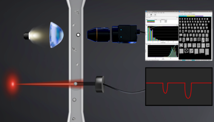
Flow imaging microscopy captures digital images of particles as they pass through a flow cell. Image-analysis software can then measure properties such as particle size, shape, transparency, and other morphological characteristics.
FIM is included in USP <1788> because particle morphology can help researchers investigate particle identity and source. Unlike membrane microscopy, FIM images particles while they remain suspended in liquid. This makes it useful for examining fragile or deformable particles, including protein aggregates and silicone oil droplets.
FIM can also provide orthogonal measurements of particle size and concentration. Because detection is based on captured images rather than light blockage alone, FIM may detect transparent particles that have limited contrast in a light obscuration measurement. Results should be interpreted according to the capabilities, settings, and validated performance of the specific method.
How Flow Imaging Microscopy Complements Compendial Testing
Flow imaging microscopy is not one of the compendial methods specified in USP <787>, <788>, or <789>. Pharmaceutical laboratories generally use light obscuration or membrane microscopy to perform the applicable compendial test.
FIM can complement those methods by supplying particle images and morphological data that they do not provide. This information may be useful when:
- A compendial test produces an unexpected particle count.
- A batch fails an applicable particulate matter test.
- A particle population changes during formulation or process development.
- A container-closure or delivery-system change introduces a new particle population.
- Researchers need to compare particle profiles across batches or stability time points.
- An investigation must assess the possible identity or source of detected particles.
For example, an increase in particle concentration may indicate a change in protein aggregation, silicone oil droplet levels, container-closure interactions, or an intrinsic or extrinsic particle source. Light obscuration records the change in particle count but cannot identify the particles responsible. Images collected using FIM can provide additional evidence to support the investigation.
FIM does not change or override the result of a compendial test. Instead, it can help researchers understand the particle population that contributed to that result.
Combining Flow Imaging Microscopy and Light Obscuration
FlowCam LO combines light obscuration and flow imaging microscopy in a single instrument. It collects LO measurements together with particle images and morphological data from the same sample flow path without requiring an additional sample aliquot for imaging.
This configuration allows laboratories to collect light obscuration data and orthogonal imaging information within the same analytical workflow. Depending on the validated application, the resulting data may support product development, process monitoring, stability studies, and investigations of unexpected particle populations.
Flow Imaging Microscopy and Silicone Oil Droplet Analysis
Many injectable protein therapies and other biotherapeutic products are supplied in prefilled syringes or other siliconized components. Silicone oil used as a lubricant can migrate into the drug product and form subvisible silicone oil droplets, also known as SiOPs.
Silicone oil droplets are analytically important because they can contribute to total subvisible particle counts but may have different origins and safety considerations from protein aggregates or foreign particles. Understanding the composition of the overall particle population can therefore provide important context during product development and risk assessment.
Flow imaging microscopy can help distinguish silicone oil droplets from protein aggregates using particle morphology. Silicone oil droplets generally appear rounded, while protein aggregates tend to have more irregular or amorphous shapes. Morphology-based filters or image-classification models can then be developed to evaluate different particle populations.
In 2025, USP published a Stimuli article explaining the rationale for a potential new informational chapter dedicated to subvisible silicone oil droplets3. Because it is a Stimuli article, it does not constitute an official USP standard and should not be used by itself to demonstrate compliance.
The Stimuli article emphasizes that no individual method fully characterizes silicone oil droplets. Light obscuration can measure particle size and concentration but cannot determine particle identity. Membrane microscopy may underrepresent silicone oil droplets because they can pass through a membrane filter. Flow imaging provides particle images and morphology, while other methods may be needed to investigate chemical identity or total silicone oil content.
Learn more about monitoring silicone oil droplets in protein therapeutics and the potential new USP informational chapter on silicone oil particles.
Applying USP <1787> and <1788> Recommendations
USP <1787> and <1788> encourage scientists to look beyond a single particle count when developing and evaluating parenteral drug products. Their recommendations support a product-specific strategy that considers the characteristics of the formulation, the particle populations likely to be present, the analytical capabilities of each method, and the purpose of the measurement.
Compendial methods remain central to demonstrating conformance with USP <787>, <788>, and <789>. Orthogonal methods such as flow imaging microscopy can add particle images and morphological information that support development studies, method comparisons, process monitoring, and investigations.
By combining particle concentration and size measurements with morphological characterization, researchers can develop a more complete understanding of protein aggregates, silicone oil droplets, and other subvisible particles in biotherapeutic drug products.
References
-
United States Pharmacopeia. USP <1787> Measurement of Subvisible Particulate Matter in Therapeutic Protein Injections.
-
United States Pharmacopeia. USP <1788> Methods for the Determination of Subvisible Particulate Matter.
-
Saggu M, Kwok SC, Narhi LO, Zhang B, Minocha S, Pombo M, Hunt DG. Addressing subvisible silicone oil droplets: industry challenges, analytical strategies, and USP's rationale for a new general informational chapter. USP Stimuli to the Revision Process. Published November 3, 2025. doi:10.31003/USPNF_S203584_10101_01.