


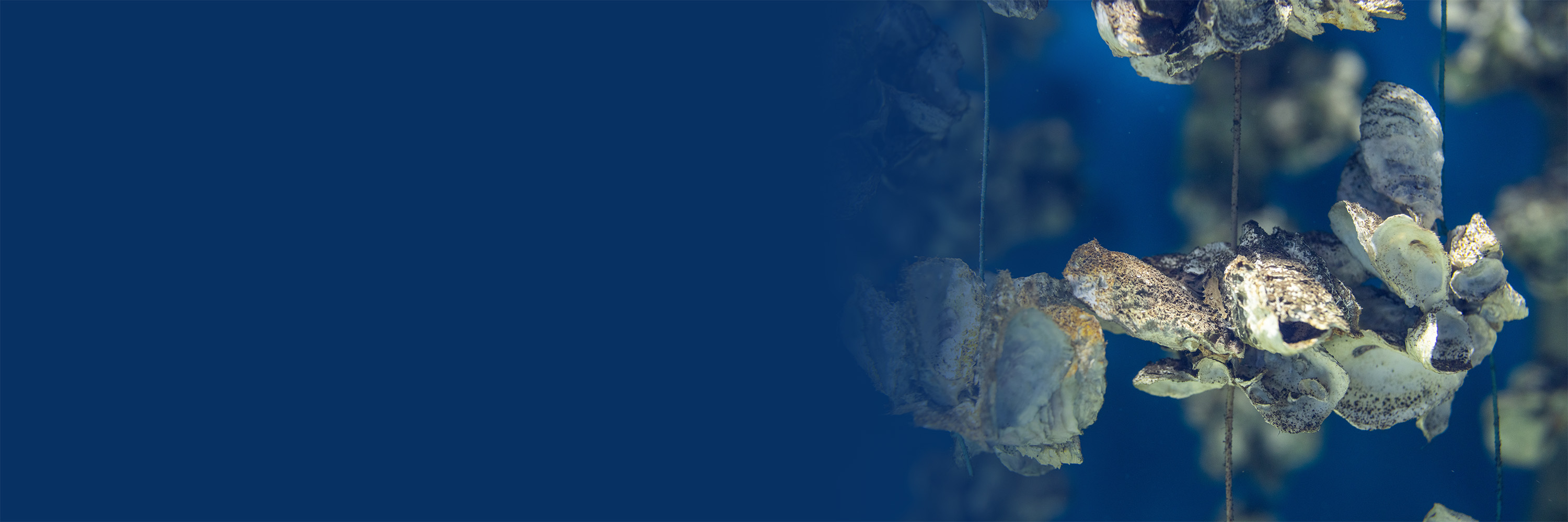
Water & Environmental
FlowCam for Shellfish Research & Aquaculture Monitoring
Automate and accelerate routine microscopy tasks to save time and increase throughput in shellfish research programs, hatcheries, finfish operations, and other aquaculture monitoring and research programs.
FlowCam’s all-in-one high-speed flow imaging microscope allows researchers and commercial operations to target microalgae and shellfish larvae on a single platform.
Whether you’re evaluating fertilization success rate and egg growth or generating a consistent, long-term monitoring record to track phytoplankton communities and harmful algal bloom events, FlowCam makes shellfish hatchery research and other aquaculture operations more streamlined.
High-Performance Shellfish Aquaculture Monitoring Capabilities
FlowCam is a powerful tool ideally suited to the needs of researchers and commercial operators alike, including shellfish hatcheries and finfish farms. Our FlowCam 8000 series, for example, is highly versatile and enables users to:
- Obtain statistically-significant count, size, and morphology data along with high-resolution images to analyze larvae, microalgae, plankton, and HABs with the same instrument.
- Quickly and reliably evaluate fertilization success rate, egg growth, development abnormalities, and contaminants.
- Rapidly count and measure larvae at several developmental stages and monitor larval transport.
- Expedite analysis of water samples to evaluate plankton availability for food supply.
- Visualize and enumerate phytoplankton to inform HAB mitigation strategies and generate long-term phytoplankton monitoring records.
"FlowCam is like a new sleuth that helps us solve mysteries in our hatchery, sometimes before we even know we have a problem!"
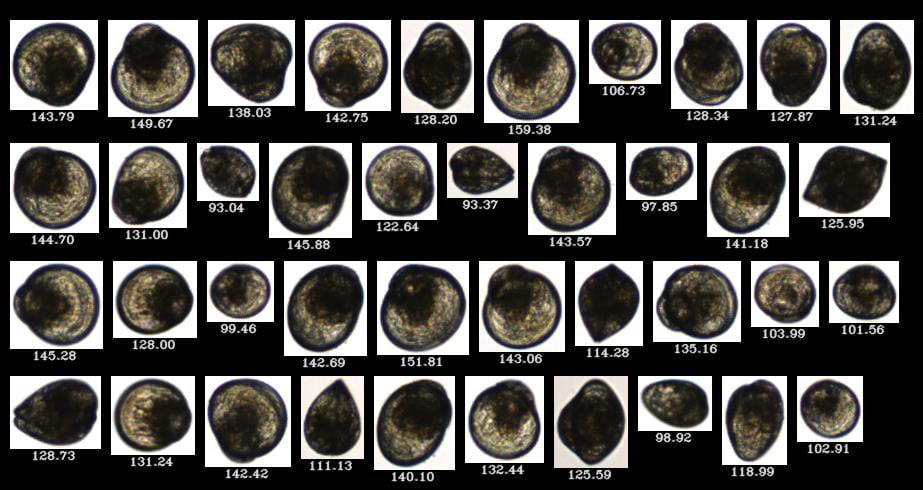
FlowCam collage of scallop spat from NOAA Milford Lab
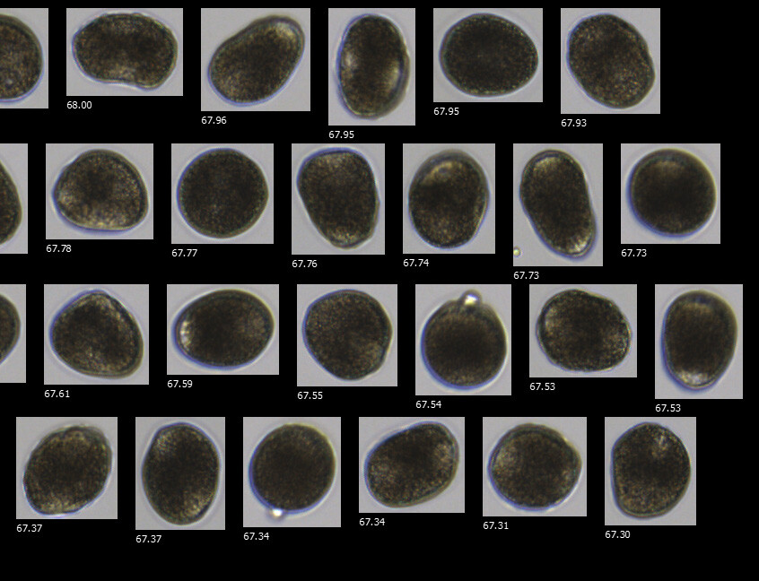
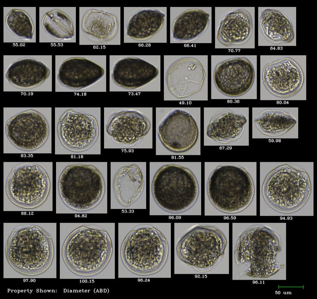
Mussel eggs with and without polar bodies imaged with FlowCam 8100 (10X, FOV100), ranging in size from 67 to 68 μm in diameter
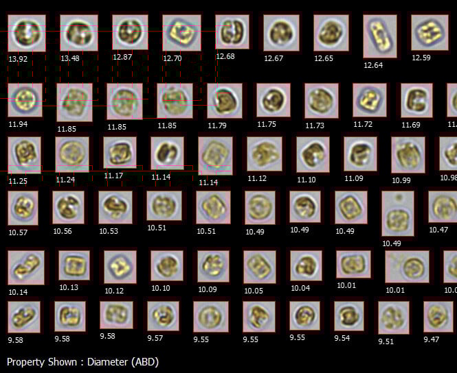
Thalassiosira weissflogii cells grown in culture by Maine's Downeast Institute shellfish hatchery, imaged by FlowCam 8100 at 20X
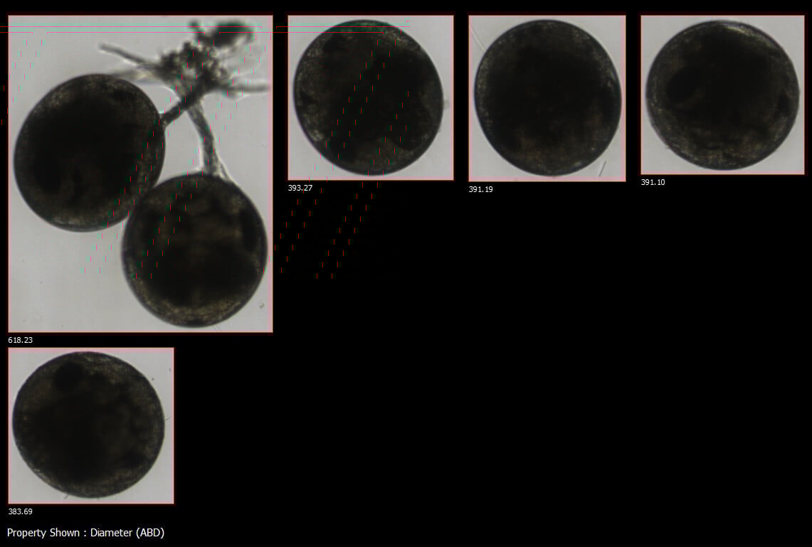
Asian shorecrab eggs imaged with FlowCam 8100 (4X, FOV600) (Source: Renee Montanaro, University of Massachusetts Dartmouth)

Chaetoceros, a diatom that can irritate fish gills, imaged by FlowCam at 10X
Ideal Applications for FlowCam Aquaculture Instrumentation & Software
Combining the basic principles of microscopy, particle counting, and image recognition, FlowCam enables rapid analysis and particle characterization for aquaculture applications as diverse as:
- Shellfish Reproduction
- Oyster Larvae Research
- Microalgae Culture Growth
- Water Quality Monitoring
- Harmful Algal Bloom Prevention
- and more...
Fluorescence-triggered imaging with the FlowCam 8400 further improves data quality. By using a laser trigger to capture images only when fluorescing particles pass through the flow cell, FlowCam 8400 minimizes noise from non-fluorescing materials, ensuring that irrelevant data is not collected.
Users can customize laser excitation to 488 nm, 532 nm, or 633 nm to target specific stains or pigments, such as FITC, phycoerythrin, Nile Red-stained lipids, or phycocyanin in cyanobacteria.
Additional Resources
 Guides
- Ultimate Guide to Flow Imaging Microscopy for Aquatic Life Sciences
- Phytoplankton Identification Image Gallery
- Flow Imaging Microscopy for Shellfish Aquaculture and Research
 Case Studies
- FlowCam Helps Scientists Track Larval Shellfish, Improve Clam Production
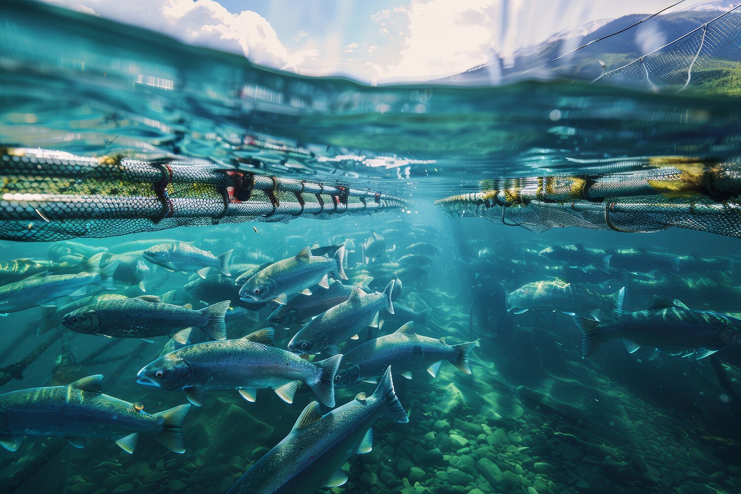
- FlowCam Assists Harmful Algae Mitigation in Salmon Aquaculture
 Videos
 Methodologies
 Blog Posts

Interested in learning more?
-
Get in Touch
Tell us about your application and particle characterization needs.
-
Have a Conversation
We're happy to set up a call to discuss your application and answer your questions.
-
Discuss Next Steps
Expand your knowledge with a seminar, demonstration, sample analysis, or obtain a quote.
