Biopharma
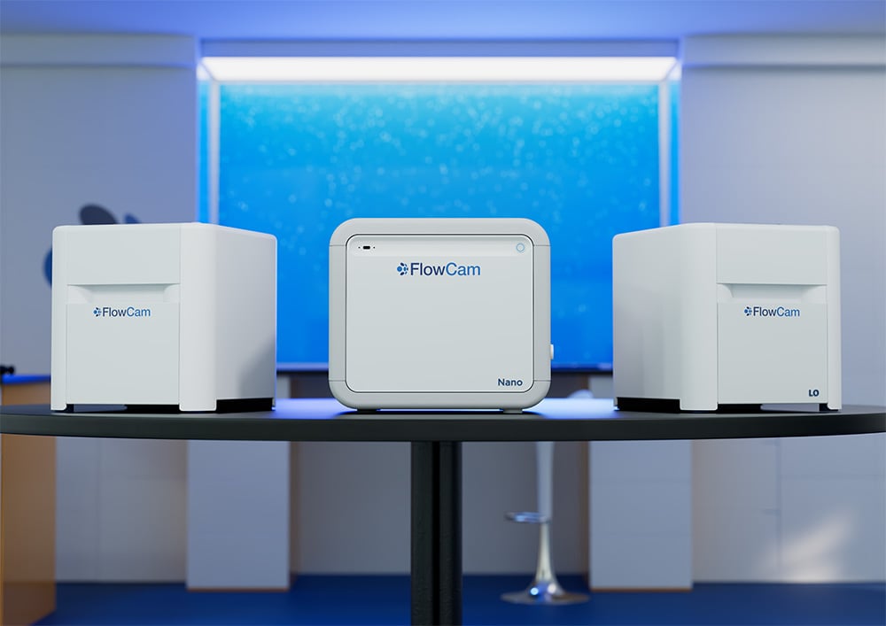
FlowCam Particle Analysis for Biopharmaceutical Development
FlowCam provides rapid, high-resolution images of subvisible and submicron particles, enabling researchers to determine concentration, size, and morphology information critical for understanding protein aggregation, cell viability, and vector integrity across protein, cell, and gene therapies.
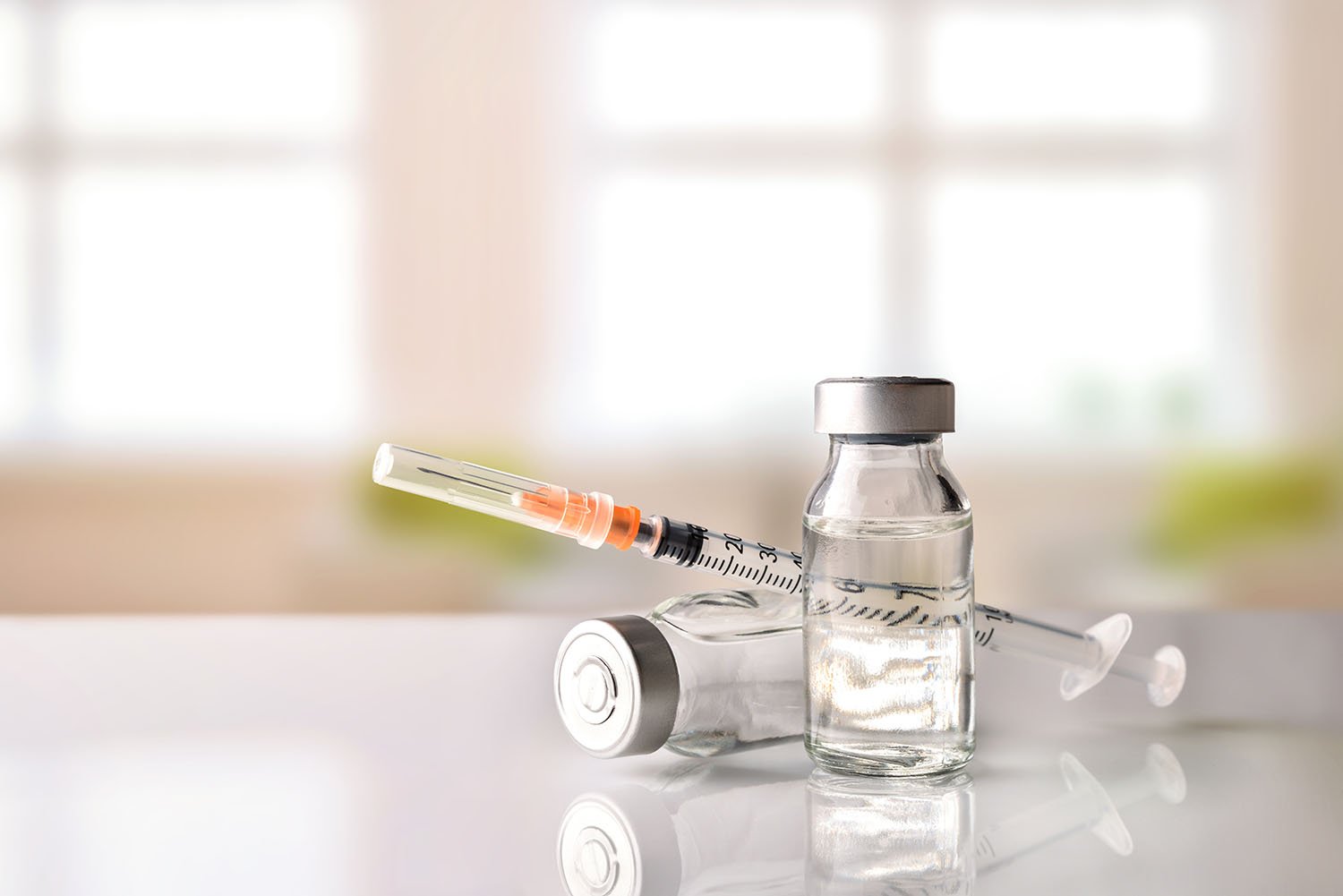
As products advance toward clinical and commercial stages, compliance with compendial particle testing requirements becomes essential. USP <787>, <788>, and <789> set standards for particulate matter in parenteral drug products. In USP <1788>, USP acknowledges the limitations of the particle testing requirements in the aforementioned chapters and recommends flow imaging microscopy (FIM) as an orthogonal method to complement traditional light obscuration and membrane microscopy. FlowCam uses FIM to deliver morphological insights that support both regulatory compliance and risk-based quality decisions.
Biopharma Application Types
Helpful Resources
Learn More About Biopharma Applications:
-
USP chapters <787>, <788>, and <789> are compendial standards issued by the United States Pharmacopeia that define particulate matter requirements for different dosage forms:
- USP <787>: applies to therapeutic protein injections, specifying limits for subvisible particles and guidance for testing methods.
- USP <788>: applies to parenteral drug products (including most biologics), requiring monitoring and restriction of particles ≥10 µm and ≥25 µm using compendial methods such as light obscuration (LO) or membrane microscopy.
- USP <789>: applies to ophthalmic solutions, setting particulate matter limits for products administered to the eye.
As products advance toward clinical and commercial stages, compliance with these standards becomes critical. USP <1788> encourages the use of orthogonal methods to capture information that LO alone may overlook. Flow imaging microscopy (FIM) is highlighted for its ability to add morphological context to particle identification and for its improved sensitivity to translucent particles.
-
While USP <788> is a compendial testing framework — and the most broadly applicable of the USP standards — USP <1788> is an informational chapter that expands on the stipulations outlined in the former. USP <1788> introduces and recommends flow imaging microscopy as an orthogonal technique to provide complementary morphological information, helping overcome undercounting and undersizing challenges with the light obscuration method during routine quality control assessments of biotherapeutics and other parenteral drugs.
-
All biopharmaceutical products contain particulate matter, as defined by the USP as mobile, undissolved particles unintentionally present in the drug product solution. Particulate matter includes subvisible particles between 2 to 100 μm in diameter, invisible to the naked eye. Subvisible particles in biologics have been associated with changes in product safety and efficacy in the clinic, including adverse reactions ranging from capillary occlusion to severe immune reactions.
-
Light obscuration (LO) measures particles by how much light they block as they pass through a laser beam. Because it assumes all particles are perfectly spherical, LO cannot provide information about particle shape, morphology, or identity.
Flow imaging microscopy (FIM), on the other hand, captures high-resolution images of every particle in the sample. This allows detailed morphological analysis and the distinction between different particle types. As an orthogonal technique to LO, FIM overcomes many of its limitations — especially for translucent or irregularly shaped particles common in biotherapeutic formulations — providing richer, more sensitive insight into particle characteristics.
-
Morphological particle analysis via flow imaging microscopy involves capturing high-resolution images to assess particle size, shape, and identity. These visual data help distinguish active pharmaceutical ingredient (API) aggregates from silicone oil droplets or other contaminants.
-
Yes, FlowCam can differentiate between aggregates of inherent API particles and contaminant particles and debris. FIM data allow users to easily and reliably detect, characterize, monitor, and control particulates in biological drug formulations.
-
FlowCam can analyze protein therapeutics (e.g., mAbs, fusion proteins); cell therapies
(e.g., CAR-T, stem cells); gene therapies (e.g., AAV, lentivirus); and other biotherapeutics requiring morphological analysis. -
FlowCam data can support regulatory filings when used within a validated method framework; many customers include FlowCam results in IND/BLA submissions.
-
FlowCam supports method validation under GMP through IQ/OQ/PQ protocols and SOP development.
-
Across models, FlowCam can detect particles between 300 nm and 1000 μm:
- FlowCam Nano: 300 nm – 2 µm
- FlowCam 8000: 2 µm – 1000 µm
- FlowCam LO: 2 µm – 70 µm
-
FlowCam instruments and VisualSpreadsheet software can reliably process samples with particle concentrations up to ~1,000,000 particles/mL. If samples exceed this concentration (whether intentionally or due to contamination), dilution is recommended to ensure accurate quantitation and avoid image overlap. For highly viscous formulations, dilution may also be necessary to enable smooth flow through the instrument’s fluidics. When diluting, it is recommended to use a buffer that is compatible with the original formulation composition to minimize artifacts (e.g., phase separation, air bubbles, or refractive index mismatches).
Additional best practices include degassing samples when possible to reduce bubble formation, avoiding harsh mixing that could introduce shear-induced aggregates, and filtering large visible particles which may clog the flow cell.
-
FlowCam 8000 and FlowCam LO can provide statistically significant results in less than a minute, with as little as 100 μL of sample.
-
Researchers and technicians seeking to capture both the required LO and the recommended FIM subvisible particle data using a single instrument and single sample aliquot might explore FlowCam LO instruments’ biopharmaceutical particle analysis solution. In addition to capturing two orthogonal subvisible particle measurements, FlowCam LO provides LO measurements that support the requirements for USP <788> and <787> testing, as well as many of the recommendations for LO measurements found in USP <1787> and <1788>. FlowCam LO is frequently used by research and development teams to help predict compendial testing results as they approach scale-up and manufacturing. FlowCam LO can be combined with automated liquid handling (ALH) for FlowCam to streamline measurements, thereby improving the repeatability of these important lot-release tests.
If your biotherapeutic formulation doesn’t necessitate LO data analysis, FlowCam 8000 enables the rapid, reliable quantification and characterization of a large volume of particles and other prevalent contaminants, including protein aggregates, cellular debris, and varying extrinsics and intrinsics. With exceptional image quality and the widest size range available on the market, this instrument is our most versatile biopharmaceutical particle size and shape analyzer. Comprehensive morphology data can help researchers and technicians improve biopharmaceutical particle analysis on a larger scale, identify hard-to-remove particulate contaminants quickly per USP standards, and eliminate them at the source. FlowCam 8000 is also compatible with ALH for FlowCam.
If you’d like to better understand how FlowCam can offer the right biopharmaceutical particle analysis instruments (and fully integrated software) for your distinct particle analysis needs, request a demo to see how our configurations work.
Need Supplies?
Find supplies and spare parts for your FlowCam instrument or ask for a quote.
Need Help?
Get technical support and application help. Request training or preventative maintenance.
Need Information?
Check out our knowledge base including white papers, application notes, technical notes, and videos.
