Using Flow Imaging Microscopy for Particle Image Analysis: How it Works
Flow Imaging Microscopy (FIM) is a rapid, high-throughput particle analysis technique that combines the benefits of digital imaging, flow cytometry, and microscopy. Beyond particle sizing and quantification, FIM provides comprehensive morphological characterization of aggregates and contaminants in biopharmaceuticals, phytoplankton and zooplankton in fresh water and marine environments, as well as advanced materials and emulsions across many industries, including food and beverage. Researchers using FIM can quickly identify, classify, and compare different particles and organisms, analyze image-based data, and share accurate, statistically robust insights.
FlowCam Improves Flow Image Analysis Capacity by Enabling:
• Direct Particle Measurements
Flow imaging microscopy enables the visualization and assessment of single particles, allowing researchers to calculate desired property values directly from individual particle images. With empirical evidence, you can advance your research, increase productivity, and ensure quality.
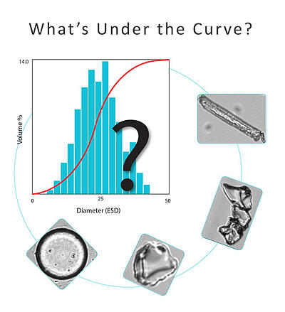
• Advanced Particle Morphology Insights
FIM also enables dynamic imaging particle analysis by providing detailed morphological characterization and allowing you to distinguish between different particle types.
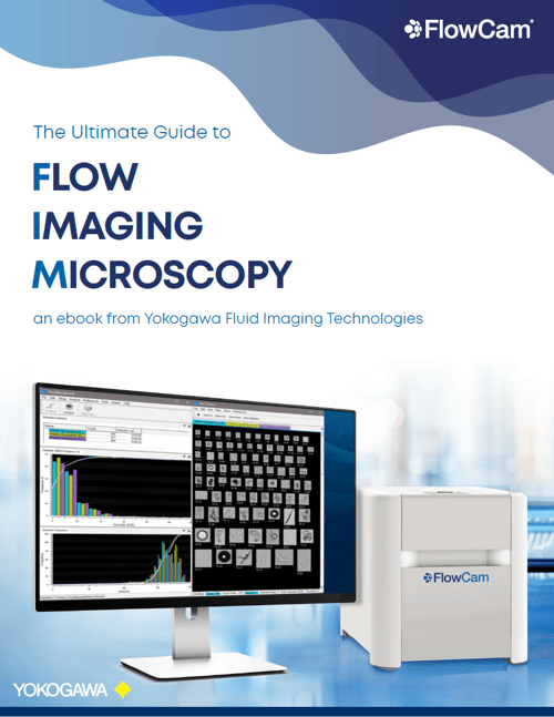
Download Our Ebook
Learn more about the benefits of FIM and how it compares to other technologies in our informative e-Book, The Ultimate Guide to Flow Imaging Microscopy

Explore Flow Imaging Microscopy Instruments from FlowCam for Particle Image Analysis
Achieve dynamic imaging particle analysis with FlowCam’s family of FIM instruments. Whether you need to detect and monitor specific types of organisms or submicron particles, FlowCam delivers the flexibility and performance to meet your needs.