Why Morphology Matters in Biotherapeutics
In biotherapeutic development—whether monoclonal antibodies, cell and gene therapies, or complex formulations—particle analysis isn’t just about counting. It’s about understanding type and source. This distinction impacts patient safety, regulatory compliance, and process optimization. Traditional methods of particle analysis, such as light obscuration (LO) and membrane microscopy (MM), fall short on particle identification. Flow Imaging Microscopy (FIM), as implemented in FlowCam, bridges this gap with morphology-based detection and high-throughput imaging.
Why Particle Characterization Matters
Subvisible particles can originate from multiple sources:
- Inherent particles: particles derived from the drug substance (e.g., aggregates of proteins, viral vectors, and lipid nanoparticles)
- Intrinsic particles: particles derived from formulation components (e.g., fatty acid particles), container-closure (e.g., silicone oil droplets), or the manufacturing process (e.g., stainless steel particles)
- Extrinsic particles: contaminant particles such as fibers or bacterial contamination
Accurate particle characterization is essential for risk assessment and regulatory compliance. As Dr. Austin Daniels, explains:
"Knowing the types of particles that are present in a sample can help scientists developing drug products identify the relevant sources of particles in their therapy formulations. That data can be helpful to get the particle content of a new formulation or drug product under control. For example, if FIM shows a lot of glass particles, it may suggest an incompatibility between the container and the rest of the formulation. This type of optimization to reduce particle content can make the product easier to manufacture and potentially perform better in the clinic."
Check out the full interview on Spectroscopy with Dr. Austin Daniels.
Why Differentiating Particle Types Matters: Regulatory Context
United States Pharmacopeia (USP) chapters such as USP <1787> and USP <1788> provide recommendations for counting, characterizing, and when possible, identifying particulate matter that could potentially be harmful to patients. These chapters apply broadly to biotherapeutics, including protein-based drugs, cell and gene therapies, and other advanced modalities.
For more information about USP standards, please refer to our white paper:
White Paper: USP Reference Standards for Subvisible Particulate Matter
FIM Enables Particle Identification and Sourcing
USP <1788> recognizes FIM as a complementary technique to MM for identifying and sourcing particulate matter, based on morphological characterization. And unlike MM, which requires buffer removal and biases against soft particle types (e.g., SO, API aggregates), FIM analyzes samples directly in their native buffer, preserving integrity and avoiding measurement bias.
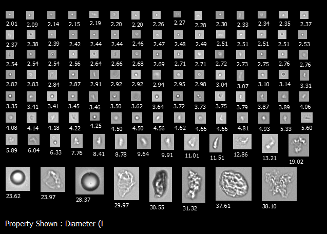
Morphological characterization with FIM instruments such as FlowCam enables particle differentiation in injectable biotherapeutics:
| Particle Type | Safety Concern | Morphology |
| Protein Aggregates | Linked to immunogenic responses | Irregular, amorphous, and textured |
| Silicone Oil Droplets | Likely less harmful, but still a product quality concern | Round and circular |
| Non-viable cells | Reduce potency and viability of the therapy | Non-circular, irregular cell shape; often fragmented |
These differences can be identified visually or through the use of automated classification tools, including AI software.
Why FlowCam Stands Out
FlowCam delivers a single, efficient solution for characterizing particle morphology as well as size and concentration—preserving particles in their native state and avoiding artifacts introduced by traditional methods such as MM. For biologics developers, morphology-based particle analysis with FlowCam isn’t just a differentiator—it’s an essential tool for ensuring safety, compliance, and efficiency.
Curious to learn more about subvisible particle analysis? Download our eBook offering insights into the latest flow imaging microscopy applications for identifying subvisible particles, including relevant USP chapters.
Download the Ebook:
Advancing Subvisible Particle Analysis: Flow Imaging Microscopy