Monitoring algae and plankton communities is central to understanding water quality, ecosystem health, and the early warning signs of harmful algal blooms (HABs). Researchers and municipalities need tools that can handle complex, mixed water samples and provide the visual detail required to distinguish organisms and effectively monitor HAB growth.
This is why many scientists search for imaging flow cytometry when working with algae and plankton. What they’re looking for is a way to combine the visual insight of microscopy with the efficiency of automated analysis without compromising accuracy.
Why Imaging Matters for Algae and Plankton Studies
Unlike mammalian cells, algae and plankton exhibit a wide range of sizes, shapes, and structures. Colonies, filaments, chains, and debris are common, and subtle morphological features often carry critical biological meaning.
For these samples, fluorescence alone is rarely sufficient. Researchers need to see:
- Colony structure and fragmentation
- Filament length and morphology
- Cell shape, size, and internal features
- The presence of debris or non-biological particles
This is where image-based analysis in flow becomes valuable, allowing large volumes of samples to be analyzed while preserving the context that only images can provide.
From Manual Microscopy to Automated Imaging in Flow
 Traditional microscopy remains the gold standard for identifying algae and plankton, but it is slow, subjective, labor-intensive, and difficult to scale. Counting and classifying organisms slide by slide limits throughput and makes long-term monitoring programs challenging.
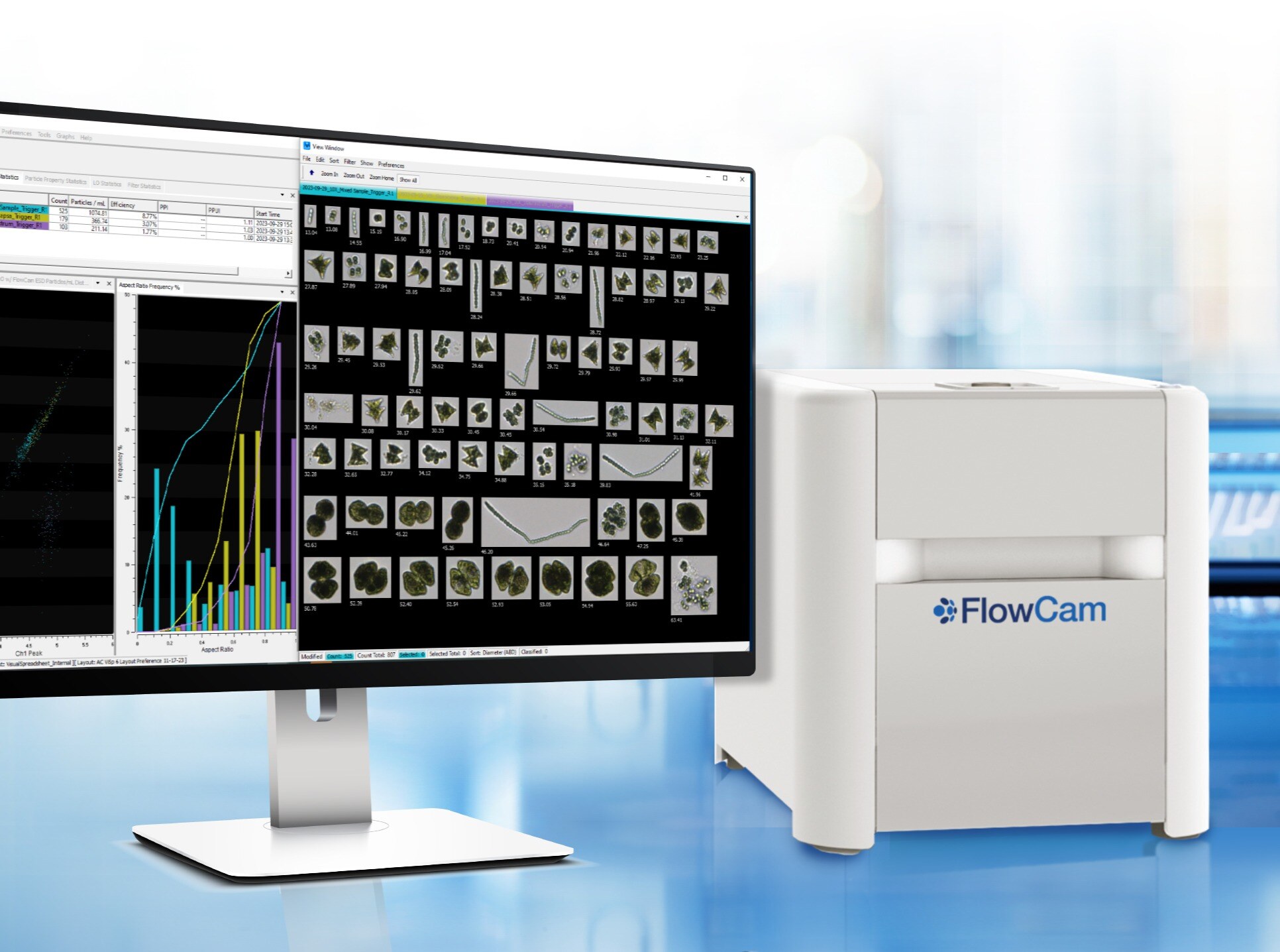
Flow-based imaging systems, such as FlowCam, address this challenge by automating microscopy. As a sample flows through the instrument, images are captured of every particle in the field of view, creating a comprehensive visual record of the sample in minutes rather than hours.
The result is high-resolution images generated fast enough to support routine monitoring, trend analysis, and large-scale studies.
Practical Advantages for HAB and Plankton Monitoring Using FlowCam
For algae and plankton researchers, the value of flow imaging microscopy instruments like FlowCam is practical and immediate:
- High-throughput quantitative analysis of heterogeneous environmental samples
- Visual confirmation of organisms, not just numerical signals
- Improved differentiation between algae, cyanobacteria, and debris supported with fluorescence triggering in FlowCam Cyano
- Archivable image datasets that support reanalysis and training of AI models
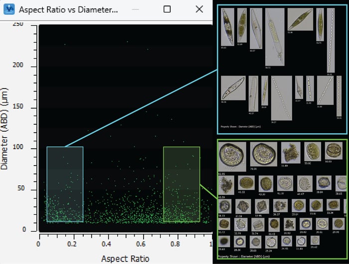
Pictured above: A marine phytoplankton assemblage (~20–80 µm), captured on FlowCam. The scatterplot shown here displays the sample data, sorted by particle diameter vs. aspect ratio, and illustrates how morphology data and images enable plankton identification and classification.
Some FlowCam models, like FlowCam Cyano, also incorporate fluorescence to assist classification, but morphology remains central—reflecting how experts actually identify organisms under a microscope.
Is FlowCam an Imaging Flow Cytometer?
In the algae and plankton research community, imaging flow cytometry has become a convenient shorthand for tools that bring imaging into flow-based analysis. While the platforms differ, FlowCam, like traditional flow cytometry, fulfills the goal of rapid characterization of particles in liquid samples, with the added advantage of capturing microscopy-quality images of every particle analyzed.
By focusing on what matters most—clear images, reliable counts, and scalable workflows—FlowCam supports the same scientific objectives driving interest in imaging flow cytometry, especially for complex environmental samples.
Seeing the Whole Community, Not Just the Signals
 For HAB monitoring, plankton ecology, and water quality research, success depends on understanding the community as a whole. Flow imaging microscopy enables scientists to move beyond indirect signals and work directly with visual data at a speed that manual microscopy simply cannot match.
To learn more about flow imaging microscopy with FlowCam, check out our Ebook, "The Ultimate Guide to Flow Imaging Microscopy".