Light obscuration (LO) is currently the compendial method for quantifying subvisible particles equal to or greater than 10 µm and 25 µm in biologic drug products. This standard is internationally recognized in the U.S. (United States Pharmacopeia or USP), European, and Japanese Pharmacopoeia. However, numerous reports have indicated that subvisible particles smaller than 10 µm also have the potential to elicit immune responses.
USP <1788> recommends the characterization of subvisible particles during the product life cycle using orthogonal methods to LO, including flow imaging microscopy (FIM).
To compare results from these analytical methods, a recent study from Japan's National Institute of Health Sciences analyzed the size and count of three different subvisible particle preparations using LO technology and two types of FIM (referred to as FI in this study) instruments: Protein Simple's MFI and Yokogawa Fluid Imaging Technologies' FlowCam. The samples were shared among 12 laboratories to compare the results across diverse settings and instruments.
It was determined that FIM may be a viable alternative to LO, as it yields many detailed morphological properties of subvisible particles. FIM has been shown to be more sensitive than LO for highly transparent particles such as proteins. FlowCam detected a relatively higher number of particles between FIM instruments than MFI.
The results are visually depicted below (Kiyoshi et al, 2019).
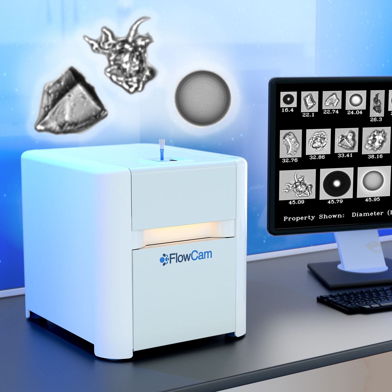
To meet USP requirements for monitoring particles in pharmaceuticals with light obscuration but also take advantage of the high-resolution images and particle morphology data offered by FIM, FlowCam LO combines flow imaging microscopy (FIM) and light obscuration (LO) into a single analytical instrument.
FlowCam LO instruments provide an innovative particle characterization solution. They allow researchers and technicians to capture both the required light obscuration and the recommended FIM subvisible particle data using a single instrument and sample aliquot.
Did you know?
MFI, short for Micro-Flow Imaging, is a term often used interchangeably with Flow Imaging Microscopy—the innovative particle analysis technology at the core of FlowCam instruments. This method provides detailed insights into particle morphology, size, and count, supporting diverse applications in biopharmaceuticals, environmental monitoring, and beyond.


