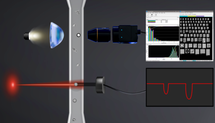
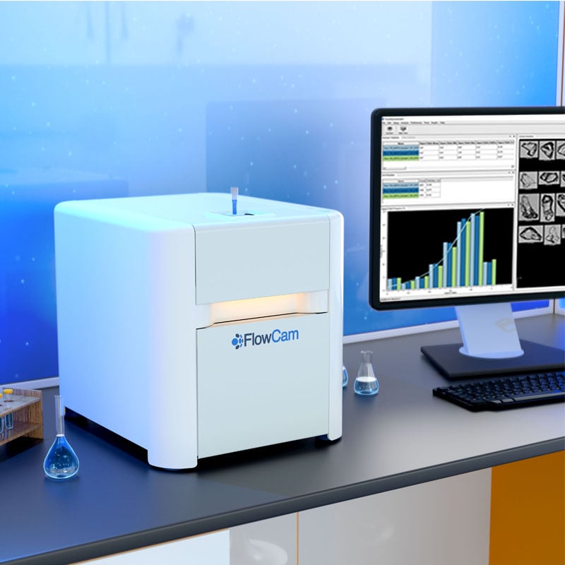





I recently joined Dr. Ashwinkumar Bhirde of the FDA and Dr. Amber Raines of Coriolis Pharma for a webinar exploring particulate matter analysis in biologic drug products. Together, we compared light obscuration (LO) and flow imaging microscopy (FIM) and discussed how orthogonal and complementary methods of analysis can support product quality assessment and particle characterization.
Watch the Webinar: "Navigating Particulate Matter Analysis in Biologic Drug Products"
Biologic drug products can contain particles from many sources, including protein aggregation, formulation components, processing, packaging, and container-closure systems. Knowing the particle count of a drug sample is important, but development teams may also need to understand what those particles look like, how the population changes, and what those changes could indicate.
Those questions shaped our webinar as Dr. Bhirde, Dr. Raines, and I explored the regulatory context for subvisible particle analysis, the complementary information available from LO and FIM, and practical examples involving stressed prefilled syringes, protein aggregates, silicone oil droplets, and formulation stability.
The recurring theme was straightforward: interpret each result according to what the method actually measures, and consider orthogonal techniques when a single measurement leaves important questions unanswered.
Light Obscuration & Flow Imaging Microscopy: Different Measurements, Different Insights
LO detects particles by measuring changes in light as particles pass through a sensing zone. It provides particle counts and estimated sizes and is an established compendial method for subvisible particle testing. During the webinar, Dr. Bhirde reviewed USP <788>, including the role of LO and membrane microscopy in testing injectable products. He also emphasized the value of orthogonal analysis, noting that orthogonal methods are recommended when additional, complementary information is needed to understand a particle population.
He explained that FIM, by contrast, captures digital images of particles in flow. In addition to size and concentration data, those images can provide information about particle morphology, including shape. This image-based view can help scientists investigate the particles underlying a numerical result and determine whether multiple particle populations may be present.
That difference is important. LO and FIM do not measure particles in exactly the same way, and results from one method should not automatically be interpreted using criteria developed for the other.
Dr. Bhirde shared examples illustrating how results can differ depending on the product and particle population being analyzed, particularly when particle properties affect how readily they are detected by LO. Watch the webinar to see the comparative data and hear Dr. Bhirde discuss what those differences mean for orthogonal particle characterization.
Why Particle Morphology Matters in Biologic Drug Product Analysis
A particle count can reveal that a sample changed, but it may not explain what changed. Particles may be inherent to a formulation, arise from processing or packaging, or enter as foreign material. These different sources can point toward very different investigations and mitigation strategies.
Prefilled syringes provide a useful example. Silicone oil droplets associated with syringe lubrication may occur alongside proteinaceous particles. A numerical count alone does not reveal which particles contributed to that result.
FIM images and morphological measurements can provide another layer of information, helping scientists group visually distinct populations and determine where further investigation may be warranted. While morphology is not chemical identification, it can help narrow the questions that need to be investigated using additional analytical techniques.
During the webinar, I shared results from three studies using FlowCam 8100 and FlowCam LO, demonstrating the value of gathering complementary information from a single sample.
Case Study: Distinguishing Protein Aggregates from Silicone Oil Droplets
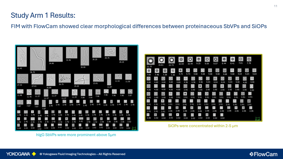
First, I shared a study using deliberately generated human IgG aggregates and silicone oil droplets to explore how FIM images and morphological measurements could be used to examine two different particle populations.
The study considered characteristics such as particle size, shape, circle fit, and symmetry and explored both the opportunities and limitations of morphology-based classification. Particle size also proved to be an important consideration when interpreting the analysis.
In the webinar, I walk through the particle images and classification approach and show how the IgG and silicone oil populations compared across these morphological parameters.
Prefilled Syringe Analysis: Reading Particle Trends Alongside Compendial Counts
I also presented a study examining spray-on siliconized prefilled syringes containing human IgG following freeze-thaw and flick stress. The resulting particle populations were evaluated using tandem FIM and LO.
The study offered an opportunity to examine the same samples through two different measurement principles. LO provided the established compendial measurement, while FIM provided images and morphological information that could help investigate the populations behind observed particle trends.
This study also reinforced an important consideration for orthogonal measurements: LO gives you the compendially relevant count data you need for compliance, whereas FIM provides the particle images required to understand what is actually behind those counts.
Watch the webinar to see how the particle populations changed following stress and how the LO and FIM measurements compared.
Formulation Stability: Using Images to Investigate Differences Between Methods
Finally, I shared a model formulation study examining variables including pH, sodium chloride, polysorbate 80, storage conditions, and time.
The study explored what scientists can learn when LO and FIM provide different views of a sample. Rather than assuming why measurements differed, we used FIM image data to investigate characteristics of the particle population and consider hypotheses that could be explored further.
This distinction is important in orthogonal analysis. Differences between methods can provide useful information, but they still need to be interpreted within the context of the product, particle population, and measurement principle.
The webinar presents the formulation comparisons, particle images, and data behind the investigation.
Building an Orthogonal Particle Characterization Strategy
Dr. Raines placed particle testing within a broader analytical toolbox. No single technique covers the entire particle size range or answers every characterization question, so the appropriate combination of methods depends on the product, stage of development, and analytical question.
For follow-up investigations, Dr. Raines discussed techniques including FTIR microscopy and SEM with elemental analysis, which can help characterize isolated particles when additional identification is required.
She also shared a real-world example demonstrating how an orthogonal investigation can move from detecting an unexpected particle population toward understanding its source and informing corrective action. Watch the webinar to hear Dr. Raines walk through the investigation and the combination of analytical methods used to identify the source.
We also discussed when to begin particle characterization during development, how limited sample volumes can affect analytical strategy, and how manufacturers can select methods appropriate to their particular drug product.
Key Takeaway: Interpret the Count, Then Investigate the Population
LO remains an established compendial method for routine subvisible particle testing, while FIM can provide images and morphological information that help scientists investigate particle populations not fully described by a count alone.
Dr. Bhirde, Dr. Raines, and I approached particulate matter analysis from different perspectives, including regulatory considerations, analytical technology, and pharmaceutical development. Taken together, the presentations illustrate why particle analysis often extends beyond asking “How many particles are present?” to questions about what those particles look like, how the population is changing, and what additional analyses may help explain why.
Watch the Webinar: "Navigating Particulate Matter Analysis in Biologic Drug Products"