


Biopharma
Advanced Organoid Analysis for Disease Modeling and Drug Discovery
Organoids are transforming biomedical research with powerful new models for studying disease and accelerating drug discovery. Yet their complexity makes analysis challenging. Variability in size, morphology, and maturation across 3D cell clusters can obscure results, compromise reproducibility, and hinder clinical translation. Traditional microscopy adds to these challenges by introducing subjectivity and lacking the throughput needed for confident, statistically robust conclusions.
FlowCam addresses these limitations with high-throughput flow imaging microscopy (FIM) and advanced morphometric analysis. By capturing thousands of high-resolution images of intact organoids in real time, FlowCam delivers precise, objective measurements of critical quality attributes – including size distribution, circularity, and shape-based metrics – providing population-level insights that support reproducibility, scalability, and translational success.
Characterize Organoids Across Research Applications
FlowCam supports a wide range of organoid research applications, including disease modeling, drug screening, toxicity evaluation, and regenerative medicine, by capturing thousands of high-resolution images and calculating detailed size and shape parameters in near real-time. This data-rich approach enables more accurate and statistically robust analysis than traditional microscopy.
With FlowCam, you can:
- Accelerate drug discovery and screening by detecting compound-driven changes such as growth inhibition and morphological degradation.
- Characterize disease models such as tumor, intestinal, and other organoid systems through detailed morphometric profiling.
- Monitor culture health and microenvironments to identify early signs of contamination, nutrient depletion, or suboptimal growth conditions through rapid imaging.
- Optimize organoid culture protocols by tracking development from early spheroid formation through maturation into complex 3D structures.
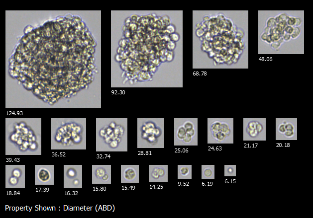
FlowCam images of human kidney organoids at various stages of assembly
Comprehensive Organoid Analysis with FlowCam
FlowCam empowers researchers to confidently assess organoid health, uniformity, and growth dynamics, accelerating the development of robust, scalable, and reproducible 3D cell culture systems for both research and therapeutic applications.
Key advantages include:
- Rapid, high-throughput capture of high-resolution images and morphometric data on thousands of intact organoids in real time.
- Wide analytical size range from 2 µm – 1 mm accommodates early spheroid formation through mature organoid structures, enabling longitudinal analysis across development stages.
- Precise quantification of key quality metrics such as size distribution, shape, circularity, and aggregate uniformity.
- Streamlined quality control workflows with objective analysis that reduces processing time from hours to minutes.

Interested in learning more?
-
Get in Touch
Tell us about your application and particle characterization needs.
-
Have a Conversation
We're happy to set up a call to discuss your application and answer your questions.
-
Discuss Next Steps
Expand your knowledge with a seminar, demonstration, sample analysis, or obtain a quote.
