In order to gain the most comprehensive understanding of particles in their samples, researchers often employ several instruments as part of their overall particle analysis scheme. Different technologies will assess different physical attributes of the particles and thereby offer scientists complementary data that are useful in validating their analyses. Even when two instruments perform essentially the same analytical technique, measurement differences between platforms may reveal physical property aspects that would otherwise go unnoticed.
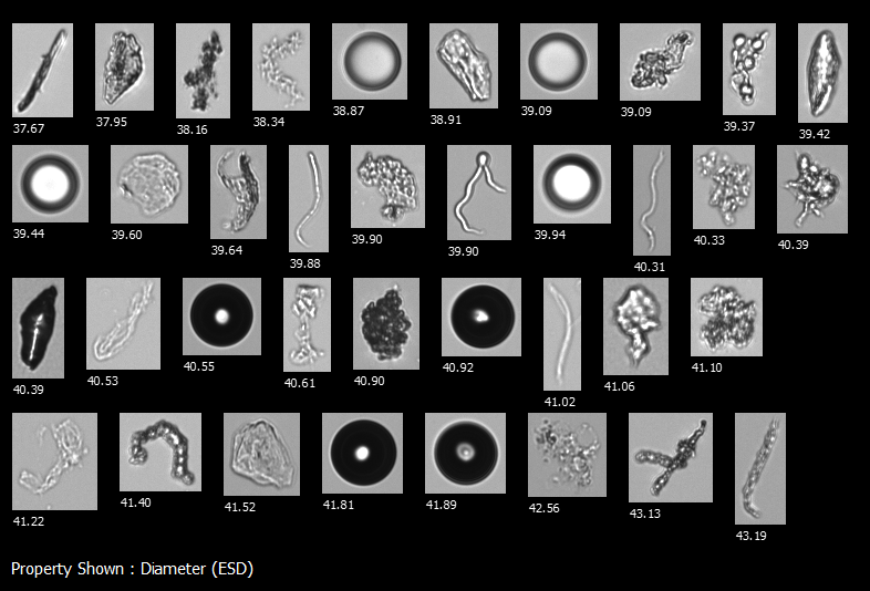
Pictured here: representative images from a biopharmaceutical sample analyzed on a FlowCam 8100 instrument.
In a recent paper published by Nishiumi et al. in the Journal of Pharmaceutical Sciences, three flow imaging microscopy instruments, FlowCam 8100, MFI 5200, and iSpect DIA-10, were compared based on their ability to size and classify common subvisible particles (SVPs) found in biopharmaceutical formulations—namely, protein aggregates, silicone oil droplets, and various lipid particles. As all three instruments offer similar particle imaging capabilities, it was of particular interest in this study to compare differences in artificial intelligence (AI) tools (like convolutional neural networks) by assessing image-based particle recognition accuracy between instruments.
Unsurprisingly, the researchers noticed differences in sizing and image analysis performance between each instrument. FlowCam 8100 performed favorably in these comparisons, especially for particle identification. AI-based image analysis tools offered higher accuracy at identifying different particle types when trained on FlowCam images than when trained on images from other platforms. This performance highlights the effectiveness of AI-based particle analysis tools like FlowCam’s VisualSpreadsheet when paired with FlowCam images, and highlights why researchers often prefer FlowCam over other flow imaging microscopy instruments for this type of SVP characterization in biopharma. Overall, the Nishiumi et al., study does a great job highlighting the strengths as well as a few shortcomings of all three flow imaging microscopy platforms for SVP analysis and illustrates the advantages of FlowCam as a flow imaging platform for your SVP analysis biopharmaceutical application.