In our previous posts, we discussed why it is critical to monitor silicone oil droplets in protein therapeutics and explored the new USP guidance emphasizing the need for more sophisticated particle characterization.
Now we're diving into the data with a new application note that demonstrates how Flow Imaging Microscopy (FIM) acts as the essential orthogonal method recommended by the USP to bridge these regulatory and technical gaps.
Download Application Note | Characterizing Subvisible Particles Using FIM
Limitations of Light Obscuration for Silicone Oil and Protein Aggregate Detection
While light obscuration (LO) has long been the compendial standard (USP <788>) for particle analysis in protein therapeutics and other parenteral drugs, it often leaves drug developers and manufacturers in the dark when it comes to the complex, low-contrast subvisible particles (SbVPs) found in modern biologics. Traditional LO works by measuring the shadow a particle casts as it passes a laser. This process is efficient for opaque, high-contrast particles, but protein aggregate SbVPs and silicone oil particles (SiOPs) are often translucent with a low refractive index. Because they don’t block much light, LO frequently undersizes or entirely misses these particles.
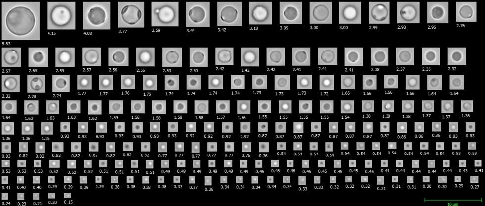
FIM, using instruments like FlowCam, captures high-resolution digital images of every particle. Instead of just a shadow, you get a full suite of morphological data—size, count, and shape—that allows you to see exactly what's in your formulation.

Differentiating Silicone Oil Droplets from Protein Aggregate Subvisible Particles
To demonstrate the effectiveness of FIM, we subjected human IgG samples to freeze-thaw stress to create protein aggregate SbVPs and used a "flick method" on siliconized syringes to generate realistic populations of SiOPs.
The results were telling:
-
Size Distribution: Both protein aggregate SbVPs and SiOPs were most prevalent in the 2–5 µm range.
-
Characterization Differences: Protein aggregate SbVPs trended larger, with ~30% of the population exceeding 5 µm, compared to less than 2% for SiOPs.
-
Visual Differentiation: While the two particle types might appear similar to LO, FIM images revealed distinct differences. SiOPs appeared as highly circular with sharp, uniform edges, while protein aggregate SbVPs were typically amorphous and translucent.
Leveraging Flow Imaging Microscopy to Identify Silicone Oil
The real power of FIM lies in its ability to turn images into statistics. Using Cohen’s d—a statistical measure of effect size—we identified which morphological properties were most effective at separating oil from protein aggregate SbVPs. While Circularity, Aspect Ratio, Circle Fit, and Circularity (Hu) all had large effects (Cohen's d values > 0.8), we chose Circle Fit as our primary indicator of roundness, since it had the lowest standard deviation for SiOPs. By building filters based on these properties, FlowCam’s software can be used to automatically classify particles in a mixed sample.
Advanced Characterization: FlowCam Nano for Submicron Silicone Oil Droplets
Given that the majority of particles observed were in the 2–5 µm size range, both the protein/hIgG aggregate SbVPs and SiOP references were also analyzed by FlowCam Nano. Its 40X oil-immersion objective revealed detailed textures—such as surface roughness on protein aggregate SbVPs and "bleb-like" features on silicone oil droplets—that are invisible at lower magnifications. This level of detail provides invaluable insight into particle behavior and the possible coalescence of SiOPs within a formulation.
Conclusion: Alignment with USP Guidance for Orthogonal Methods
 As biologics continue to grow as a class of medicine, the need for independent verification of Critical Quality Attributes (CQAs) has never been higher. FIM doesn't just provide a count; it provides the visual evidence necessary to identify the root causes of particle formation and ensure patient safety. By integrating FIM into your analytical workflow, you move beyond the limitations of LO and align with the latest USP stimuli guidance for comprehensive particle characterization.
Download the full application note, Characterizing Subvisible Particles Using Flow Imaging Microscopy: Aligning with New USP Guidance of Silicone Oil Droplets, to see the detailed data and learn how to optimize your particle identification workflow.