The uniformity of printer toner particles affects the distribution of charge the particles hold and as a result, can affect the image quality of the printed materials. Particle analysis can be used to determine shape, size, circularity, and material uniformity of printer toner particles during and after production.
Particle analysis involves taking a sample of a substance and analyzing the individual particles of the sample. The basic goal is to determine the constituents of a mixture and to differentiate between particles in the sample. Typical measurements of particles that are of interest include: particle size distribution, particle count, various measurements of particle shape, and particle concentration.
Volumetric Partle Analysis Techniques
Rapid particle processing requirements have led to the development of several high-volume, indirect measurement techniques such as the Coulter counter, laser diffraction, light obscuration, and dynamic light scattering.
There are advantages and disadvantages to volumetric particle analysis. The advantages include the ability to: rapidly determine size and count, obtain statistically significant data, create particle distribution graphs, and procure detailed particle statistics. The primary disadvantage of these methods is that they assume that particles are spherical. The analysis is limited to count and size, and shapes cannot be distinguished.
Flow Imaging Microscopy
While volumetric methods are sufficient to calculate particle size, when shape and/or morphological data are needed, a more in-depth analysis is required to truly characterize a particle.
Flow Imaging Microscopy (FIM) combines the benefits of manual microscopy with those of volumetric techniques. Microscopic particle measurements are taken from images quickly enough to produce statistically significant results. Additionally, multiple measurements are taken for each particle, thereby providing the detailed information often needed for a thorough particle analysis. The addition of specialized software provides sophisticated post-processing of data as well as the ability to see real images of your particles, giving you an in-depth understanding of your sample.
How it Works
Flow imaging microscopy uses digital images to measure the size and shape of each particle. Essentially, the operator in traditional microscopy is replaced by a computer to extract the information from the images.
The sample containing the particles streams through a flow cell past microscope optics. Thousands of particle images are captured per second. To capture sharp images of moving particles, they are “frozen” in space using a strobed illumination source combined synchronously with a very short shutter speed.
As each frame of the camera’s field of view is captured, the software, in real-time, extracts the particle images from the background and stores them.

In an imaging-based system, particle measurements are made directly from the image of the particle. Since the system’s optics are fixed and the magnification is known, distance measurements on the image can be directly converted to real distance measurements on the object. No generalizations are made about a particle’s shape. Plus, you can view the image to ensure that the data is being properly interpreted.
Common measurements include: equivalent spherical diameter; area based diameter, length, width, and aspect ratio; area and volume; circularity and elongation, edge gradient; intensity, average intensity, and sigma intensity; transparency, and many more. The FlowCam® flow imaging microscope calculates more than 40 properties for each particle.
Watch a video: How does FlowCam work?
Flow Imaging Microscopy vs. Laser Diffraction
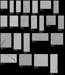
 These images are from a customer using laser diffraction to characterize the particles in their fluids. They had a sense that the data they were getting was not accurate. Laser diffraction results indicated that there were no particles larger 65 µm in their samples.
They decided to test FlowCam, and when doing so, found not only many irregularly-shaped particles in their samples, but also many contaminant particles greater than 90 µm that were missed entirely by laser diffraction.
The technology used in laser diffraction (like other non-imaging technologies) converts all particles to an equivalent spherical diameter (ESD). In certain cases, long fibers would be counted as small spheres. And in other cases, translucent particles were missed entirely. Laser diffraction is not a reliable source for registering and sizing particles with a low aspect ratio.
FlowCam for Printer Toner Analysis
Image analysis enabled by flow imaging microscopy is crucial to the ability to measure particle circularity, one of the principal properties relevant to quality control analysis in the manufacturing of printer toner. The combination of particle size and shape monitoring, as made possible with FlowCam, enhances the quality control process essential to printer toner manufacturers.
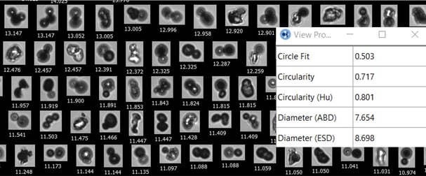
With FlowCam's software, VisualSpreadsheet, filters can be created to look for toner aggregates, by filtering for low circular fit and circularity properties.
Low circularity toner particles:
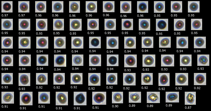
High-circularity toner particles:

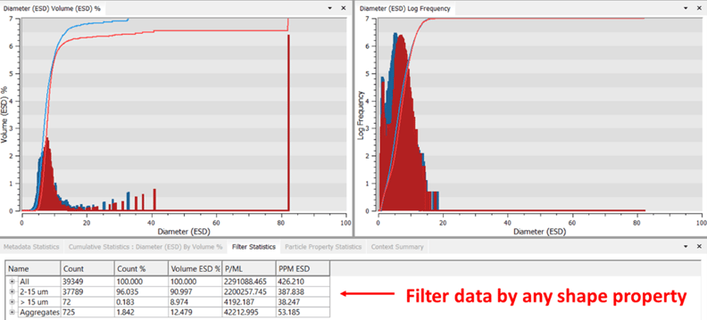
VisualSpreadsheet also allows you to see not only the whole particle size distribution (PSD) but also to analyze specific ranges and outliers separately, which can be done with software filters based on morphology characteristics.
Summary
Image analysis enabled by Flow Imaging Microscopy is crucial to the ability to measure particle circularity, one of the principal properties relevant to quality control analysis in the manufacturing of printer toner. Other high-volume particle analysis techniques are able to determine particle size, but since they assume that all particles are spheres, they do not allow for particle shape analysis.
 The combination of particle size and shape monitoring, as made possible using FlowCam, enhances the quality control process essential to printer toner manufacturers. Implementing a QA/QC filter can help assess the general quality of each analyzed sample. FlowCam is well adapted for a broad range of applications that necessitate rapid, quantitative results. The instrument has an intuitive design that enables an operator to conduct on-site testing of a product with conformational data, statistics and reporting.
In our application note, "Printer Toner Quality Assurance with FlowCam", we demonstrate that the FlowCam can be used for rapid quality control characterization of printer toner.