The occurrence and severity of harmful algal blooms are on the rise due to climate change. This trend necessitates the development of a reliable and scalable approach for public safety and conservation agencies to swiftly detect and quantify colonies of cyanobacteria. FlowCam is an established technology that facilitates the identification of phytoplankton at the genus level, and sometimes species level, and estimates the abundance of individual cells by combining the benefits of flow cytometry and light microscopy. FlowCam Cyano offers an even faster approach to classification by using a 633 nm laser to detect the presence of phycocyanin and chlorophyll, thereby identifying cyanobacteria and other algae in water samples automatically.
FlowCam is being used globally to estimate the cell abundance of colonial cyanobacteria, including Microcystis sp., a globally pervasive genus of blue-green algae, which is frequently seen in source water systems and recreational lakes. These cell colonies can range in size from a few microns up to several thousand microns in diameter. When analyzing large colonies, researchers may worry about losing part of the sample or not being able to capture a quality image due to the colony’s size and density. Dense colonies superimpose cells in the image, making it easy to underestimate cell counts, while large colonies can make it challenging to determine which objective magnification is best used to image the sample.
What is the Preferred FlowCam Methodology for Analyzing Microcystis Colonies?
 There are multiple possible methods of processing a Microcystis sample, and the method that's right for you will vary depending on the density and concentration of the colonies in your samples.
Our technical note, Estimating Cell Counts for Colonial and Filamentous Cyanobacteria, describes a peer-reviewed procedure, developed by the California Department of Water Resources (USA), that is commonly used to count cells of colonial cyanobacteria, including Microcystis and Dolichospermum (Anabaena). This strategy parallels the one used with manual microscopy, but leverages the larger and more statistically significant data sets provided by FlowCam.
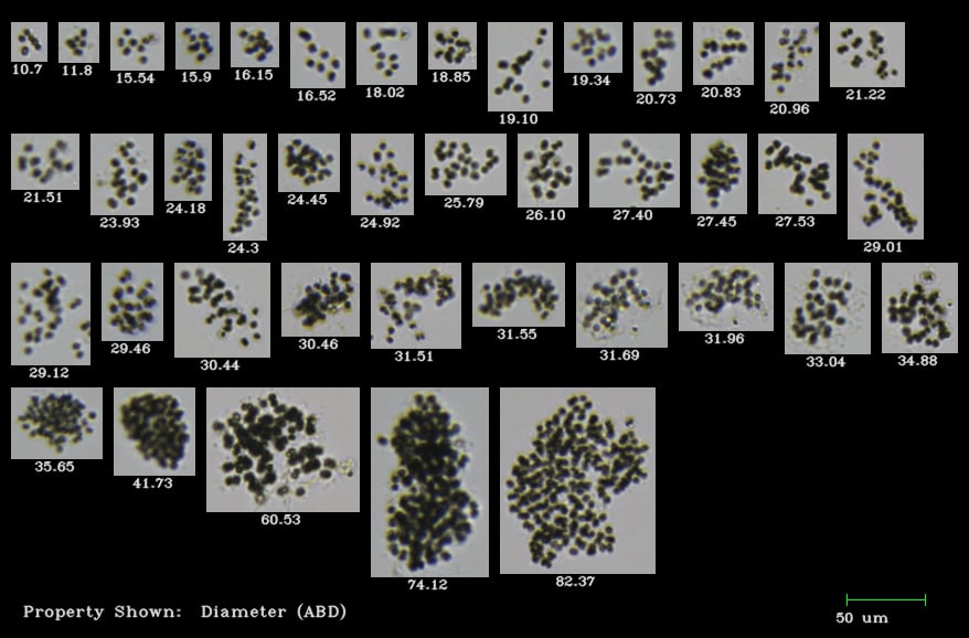
A Novel Method for Cell Counting of Microcystis Colonies
In a study published in Environmental Engineering Research, Park et al. present their own method for cell counting Microcystis colonies using FlowCam. Researchers from Korea Water Resources Corporation, University of Central Florida, and Kyungbook National University developed this three-dimensional image processing method using an algorithm to count colonial Microcystis cells.
First, Microcystis colonies within a water sample were identified with FlowCam using a local FlowCam image library of Microcystis colonies collected from rivers and reservoirs in South Korea. Second, the total sum of the areas of the Microcystis colonies within the water sample was obtained using FlowCam. Then, a model algorithm developed in this study (the relationship between the number of cells vs. the 2D surface area of the algal colonies) was applied to estimate the total number of individual cells of Microcystis sp. within the water sample. The accuracy of the model algorithm in counting the total number of individual cells in Microcystis colonies within the water sample was then evaluated by comparing the result FlowCam results with the results from a conventional microscopic method for algal cell counting.
Additional Tips and Tricks for Analyzing Microcystis Colonies with FlowCam
To ensure the most accurate and efficient analysis of Microcystis colonies using FlowCam, it’s important to tailor your sample preparation and analysis techniques to the unique characteristics of your samples. The following tips and best practices can help you overcome common challenges—such as clogs, dense colonies, and image quality issues—and improve the reliability of your results:
- Microcystis will change size/shape according to the environment that it’s in. It’s common to see a 300 µm colony pass through a 100 µm-deep flow cell. Dilution with de-ionized water can facilitate smoother processing if the colonies are large, ensuring that the colonies are dispersed enough to pass through the flow chamber one at a time. Microcystis is more forgiving than filamentous organisms like Aphanazomenon, which are more likely to clog when they cluster together.
- Samples can be filtered with mesh to remove large sediments/colonies and eliminate the potential for a clog. If you find that clogs are occurring frequently, we recommend the following:
- Analyze a second sample at 4X with a larger flow cell (300 or 600 µm deep).
- Rinse the mesh onto a slide and briefly scan it under a microscope to visually confirm if the data resembles your FlowCam data. This is common since the microscope is an excellent tool for qualitative (as opposed to quantitative) data collection and requires little additional time for the operator.
- For dense samples that can occur during an algae bloom, the operator can sonicate or shake the sample to disperse the colonies so the cells can be counted individually.
- Cell counts do not indicate toxicity; instead, they are a low-cost, fast procedure used to quantify risk. Toxin tests can become extremely expensive, so it’s cost- and time-efficient to identify if toxin-producing organisms are present and significantly abundant.
Learn how FlowCam is used to Monitor Harmful Algal Blooms
References:
Park, J., Kim, Y., Kim, M., and W. H Lee, 2018, A novel method for cell counting of Microcystis colonies in water resources using a digital imaging flow cytometer and microscope. Environmental Engineering Research, doi: 10.4491/eer.2018.266.