Microcystis is a genus of Cyanobacteria containing species capable of producing toxins called microcystins, which are harmful compounds that pose significant health risks to humans and wildlife. These toxins can lead to various adverse effects, including liver damage and skin irritation, and are a major concern for water quality management.
Microcystis can be found in eutrophic waterbodies worldwide, thriving in nutrient-rich environments where they can form dense blooms. These blooms often appear as thick, green mats on the water's surface, which can disrupt aquatic ecosystems and lead to oxygen depletion. It is so ubiquitous that when someone hears the terms “Cyanobacteria” or “harmful algal bloom” (HAB) about a lake or reservoir, Microcystis is almost always part of the conversation. This is because it is one of the most common and well-known contributors to HABs, which can have severe ecological and economic impacts. Knowing whether Microcystis is present in a water body is a key factor in determining the level of risk for a HAB and/or potential toxin event. Monitoring its presence helps assess the potential for toxin production and implement timely management strategies to protect public health and the environment.
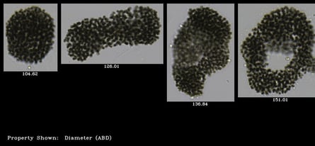
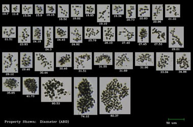
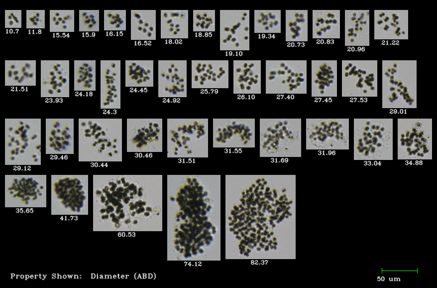
Pictured here: examples of large Microcystis sp. colonies, imaged by FlowCam Cyano
Quantifying Microcystis can be challenging due to how it forms colonies. Several techniques can be used to detect Microcystis, including flow imaging microscopy. This method combines the principles of light microscopy and flow cytometry to capture images of particles and organisms in a fluid stream.
FlowCam Cyano takes this technology a step further by automatically differentiating between fluorescence-emitting Cyanobacteria and other algae. Polly Barrowman, our Water Quality Monitoring Specialist, presented at the 2023 Harmful Algal Bloom Symposium hosted by the University of Wisconsin.
In her presentation, Polly covers the following topics:
- An overview of flow imaging microscopy for water quality monitoring
- A summary of the key studies that reflect how flow imaging microscopy has historically been used to study Microcystis
- A novel method for cell counting of Microcystis colonies in water resources using a digital imaging flow cytometer and microscope - Park et al 2019, Environmental Engineering Research
- A quantitative protocol for rapid analysis of cell density and size distribution of pelagic & benthic Microcystis colonies by FlowCam - Wang et al 2014, Journal of Applied Phycology
- Semi-automated classification of colonial Microcystis in mesocosm experiment reveals high heterogeneity during seasonal bloom - Mirasbekov et al 2021, Scientific Reports
- A reflection on the benefits and limitations of flow imaging microscopy for studying Microcystis and other colonial organisms like it
Download the presentation here:
A Review of Recent Studies of Microcystis Using FlowCam
Learn more about FlowCam for water quality monitoring on our website.