Cell-based medicinal products (CBMPs) are gaining importance as therapeutic treatments for many life-threatening diseases. This study was designed to develop a method to characterize CBMPs using flow imaging microscopy (FIM) for the detection, quantification, and characterization of subvisible particulate impurities. Recently, FIM has been applied to study cell viability and confluency in cell culture as well as quality of CBMPs. Comprehensive particle characterization can be limited when using the morphological parameters derived from the embedded software due to the complexity of CBMPs, which may consist of multiple particulate populations of highly heterogeneous morphologies.
Deep learning for image analysis is an alternative approach, offering more insight into the collected data and potentially allowing for better discrimination of particle populations. The increasing computational power and advancements in algorithms for pattern recognition have made the deep learning methods, such as CNNs, useful tools in the biopharmaceutical industry.
Jurkat cells and Dynabeads were used to represent cellular material and non-cellular particulate impurities, respectively. This study demonstrated that FIM assisted with CNN is a powerful method for detecting and quantifying cells. 
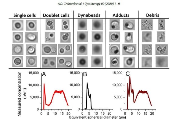
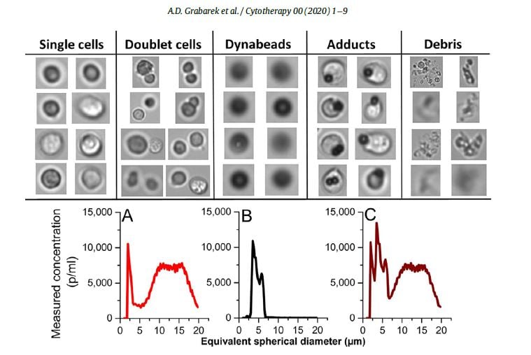
Pictured above, representative images of each population class obtained by using the FlowCam.
By using CNN, the authors experienced a 50-fold reduction in misclassification rates, compared with the use of only the output parameters from the FIM software. They conclude that CNN-assisted FIM is a powerful method for the detection and quantification of cells, beads and other subvisible process impurities potentially present in CBMPs.
Read the full paper here as published in Cytotherapy, International Society Cell & Gene Therapy.
Particulate Impurities in cell-based medicinal products traced by flow imaging microscopy combined with deep learning for image analysis.
A.D. Grabarek, E. Senel, T. Menzen, KH Hoogendoorn, K. Pike-Overzet, A. Hawe, W. Jiskoot


