


Dynamic image analysis (DIA) is a powerful technique for characterizing particles in motion, enabling real-time measurement of size, shape, and morphology. By capturing high-resolution images of particles in motion, DIA preserves sample integrity while delivering a more complete and representative view of particle populations than traditional static methods.
Within this broader category, flow imaging microscopy (FIM)—the technology used by FlowCam—is a specific and widely adopted implementation of DIA that combines imaging, automation, and quantitative analysis for particles in a fluid suspension.
Together, the terms DIA and FIM describe the same fundamental capability: high-throughput, image-based particle characterization in a flowing system – and can be used interchangeably.
How Dynamic Image Analysis (Flow Imaging Microscopy) Works
DIA, often referred to as FIM, integrates microscopy with automated, high-throughput analysis:
Sample Introduction
A liquid sample containing particles or microorganisms is added to the sample port, either manually or via automated sampling, and drawn into the microfluidic system by a high-precision pump.
Flow and Image Capture
As particles pass through the optical flow cell imaging region, a high-speed camera captures sharp images using synchronized illumination to eliminate motion blur.
Image Processing and Measurement
Software isolates each particle and calculates quantitative morphological data directly from the images. Particle properties, including size, shape, and light interaction properties such as transparency and intensity, are thus determined directly from calibrated and fixed optical settings without mathematical assumptions.
Data Visualization and Analysis
Users can review individual particle images alongside quantitative data, apply statistical filters, and classify particles by morphological properties — enabling deeper image-based insight than size-only techniques.
Dynamic Imaging: Why it Matters?
Traditional particle analysis methods each have limitations:
-
Static microscopy provides detailed images but is slow, manual, and prone to user bias
-
Laser diffraction delivers size distributions, but without imaging, it lacks shape and morphological detail
-
Flow cytometry relies on inherent fluorescence or labeled biological markers to detect and count particles, but offers limited structural insight.
DIA, especially in the form of FIM, bridges the throughput and morphological gaps of traditional systems. It enables rapid, automated analysis of thousands of particles while capturing both size and morphology in a single pass. The combination of throughput and detail makes FIM uniquely valuable across industries.
Key Applications of Dynamic Image Analysis with FIM
Because particle characteristics directly impact product performance and safety, DIA plays a critical role in multiple fields:
Biopharmaceuticals
Detects and characterizes subvisible particles (e.g., protein aggregates, silicone oil droplets, etc.), supporting drug safety and formulation stability
Aquatic Research
Enables identification and monitoring of plankton and harmful algal blooms, supporting environmental analysis
Materials Science
Provides detailed morphology of particles in advanced materials, including powders, polymers, and additive manufacturing materials
Food and Beverage
Helps monitor emulsions, suspensions, and ingredient consistency
FlowCam: Bringing DIA to Life Through Flow Imaging Microscopy
FlowCam systems are purpose-built implementations of dynamic image analysis through flow imaging microscopy. By combining high-resolution imaging with automated analysis, FlowCam enables rapid, reproducible particle characterization across a wide range of applications.
With the ability to analyze thousands of particles in minutes, FlowCam delivers both quantitative data and visual confirmation, helping users move from sample to insight more efficiently. Different models support specialized workflows, from biopharmaceutical analysis to aquatic research.
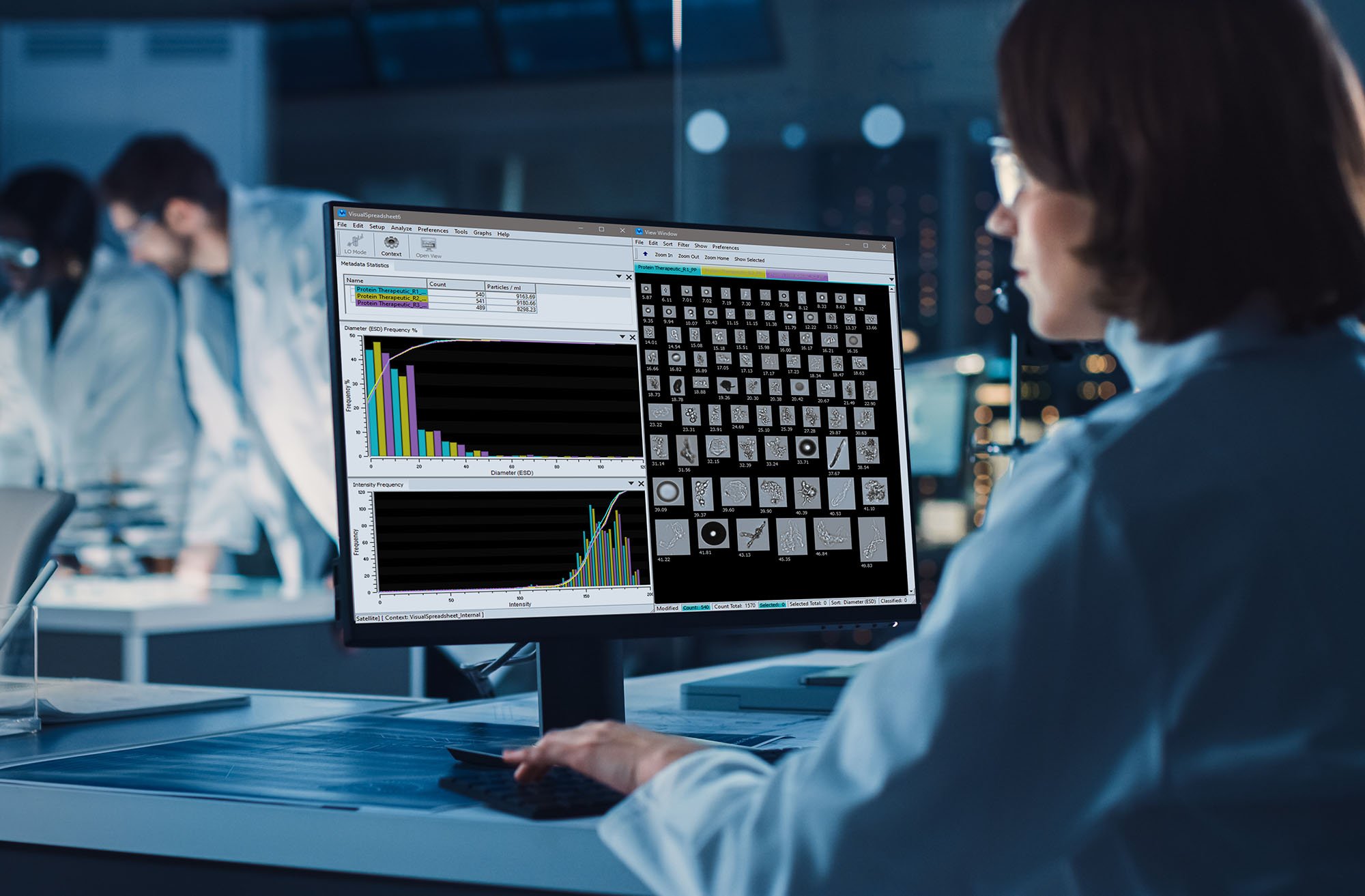
Paired with VisualSpreadsheet software, FlowCam allows users to sort, filter, and classify particles with ease—turning large image datasets into actionable information.
Bringing it all Together
Dynamic image analysis is an overarching methodology for characterizing particles in motion.
Flow imaging microscopy is how DIA is realized for particles in liquid suspension.
FIM with FlowCam translates the power of DIA into a robust, application-ready solution for liquid particle suspensions across a wide array of applications from advanced materials, aquatics, biopharmaceuticals, and beyond.
Ready to explore further?
Download our eBook, The Ultimate Guide to Flow Imaging Microscopy, to learn how FlowCam can support your particle analysis workflow:


