Flow Imaging Microscopy (FIM) is finding its pulse in human health applications through our collaboration with the MaineHealth Institute for Research (MHIR). We teamed up with scientists from the Myocardial Biology and Heart Failure (MBHF) research lab in the Center for Molecular Medicine (CMM) to demonstrate the value of FlowCam Nano in translational research. Our MBHF partners are especially interested in the changes that occur in the particle composition of biological fluids in response to cardiac stress, and they are asking questions that are aimed at moving basic science discoveries into clinical practice. With FlowCam Nano, we hope to help them find answers that can bridge that bench-to-bedside gap and benefit human health.
Virtually all cells release extracellular particles of various shapes and sizes into biological fluids (including blood, saliva, sputum, tears, and cerebrospinal fluid) due to normal cellular processes.1 Extracellular vesicles (EVs) are a class of extracellular particles that have a spherical structure composed of a lipid bilayer. EVs include exosomes (size range of 30-100 nm), microvesicles (MVs), (size range of 100-700 nm), and apoptotic bodies (sizes of up to 5 µm) – the latter of which may contain organelles and nuclear components.2 After an event like cardiac arrest, changes in EVs sizes, morphologies, concentrations, and size distributions can reveal the severity of tissue damage in the heart and/or brain and thus could be used for diagnosis, monitoring, and even the development of new therapies for various cardiovascular conditions.3,4
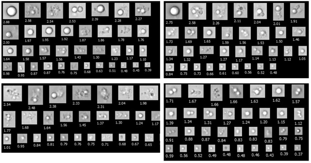
Characterizing particles in the 300 nm to 2 µm range in human plasma is no small task, but the dynamic imaging capabilities of FlowCam Nano make it possible. Using a 40X objective, the instrument captures high-resolution images of submicron particles, while VisualSpreadsheet software creates a catalog of morphological features that can be culled for meaningful trends in particle properties. So far, FlowCam Nano has given our MBHF collaborators a close-up view of submicron particles in biological fluid samples. We look forward to furthering our detailed analysis already in progress and adding value to the wealth of metadata and molecular information they have accumulated over the years.
The four collages below show FlowCam Nano images of extracellular vesicles obtained from four different biological fluid samples.
Central to MHIR’s research success is the collaborative network it fosters among Maine Medical Center physicians and other investigators in the region. We are excited about our collaboration with MHIR and the opportunity to explore the biological fluid imaging capabilities of FlowCam Nano.
When asked what the big picture of their research mission is, our MBHF collaborators, Sergey Ryzhov, MD, Ph.D., and Joanne Taylor deKay, MS, said quite simply: “To help people rejuvenate their aging hearts and improve their overall cardiovascular health." Perhaps together, we can.
Learn more about how FlowCam Nano can provide early detection of aggregation in formulations.

References
- Neri T, Celi A, Tinè M, Bernardinello N, Cosio MG, Saetta M, Nieri D, Bazzan E. The Emerging Role of Extracellular Vesicles Detected in Different Biological Fluids in COPD. International Journal of Molecular Sciences. 2022; 23(9):5136. https://doi.org/10.3390/ijms23095136
- Pulliero A, Pergoli L, LA Maestra S, Micale RT, Camoirano A, Bollati V, Izzotti A, DE Flora S. Extracellular vesicles in biological fluids. A biomarker of exposure to cigarette smoke and treatment with chemopreventive drugs. J Prev Med Hyg. 2019 Dec 20;60(4):E327-E336. doi: 10.15167/2421-4248/jpmh2019.60.4.1284. PMID: 31967089; PMCID: PMC6953455.
- deKay JT, May TL, Riker RR, Rud J, Gagnon DJ, Sawyer DB, Seder DB, Ryzhov S. The number of circulating CD26 expressing cells is decreased in critical COVID-19 illness. Cytometry A. 2023 Feb;103(2):153-161. doi: 10.1002/cyto.a.24547. Epub 2022 Mar 16. PMID: 35246910; PMCID: PMC9087143.
- Fu, S., Zhang, Y., Li, Y. et al. Extracellular vesicles in cardiovascular diseases. Cell Death Discov. 6, 68 (2020). https://doi.org/10.1038/s41420-020-00305-y