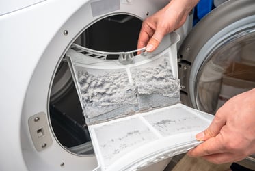






Microplastics have become a growing concern for researchers, regulators, and water utilities worldwide. As plastic waste breaks down in the environment, microscopic particles can accumulate in rivers, lakes, oceans, and drinking water sources. While advanced techniques such as FT-IR and Raman spectroscopy can identify polymer types, they often require significant analysis time, limiting sample throughput.
This challenge has created a need for rapid screening methods that can help laboratories identify samples of concern before committing resources to more detailed polymer characterization. Flow imaging microscopy (FIM) offers a promising solution by combining particle imaging, enumeration, and morphology measurements in a single workflow.
Why Rapid Screening for Microplastics Matters
Plastics have endowed society with remarkable advancements from medicine and transportation to electronics and consumer goods. Their low cost, versatility, and durability have fundamentally reshaped modern life. However, the persistence of plastic litter in the world’s oceans, lakes, and land-based resources is threatening natural habitats—endangering animal life, the food chain, and public health.
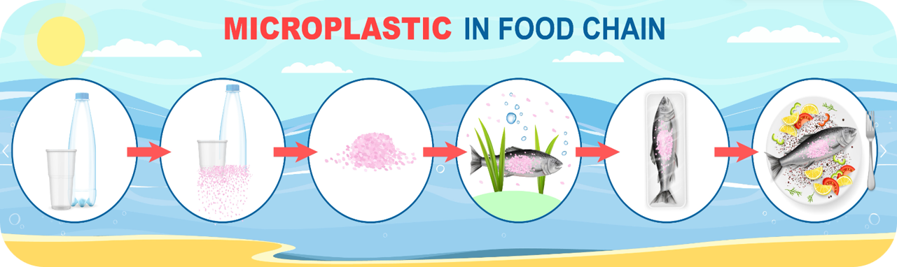
Microplastics are increasingly being monitored because of their prevalence in aquatic environments and their potential to act as carriers for contaminants such as algal toxins and pharmaceutical compounds. Researchers and water utilities often need to analyze large numbers of samples, making speed and efficiency essential.
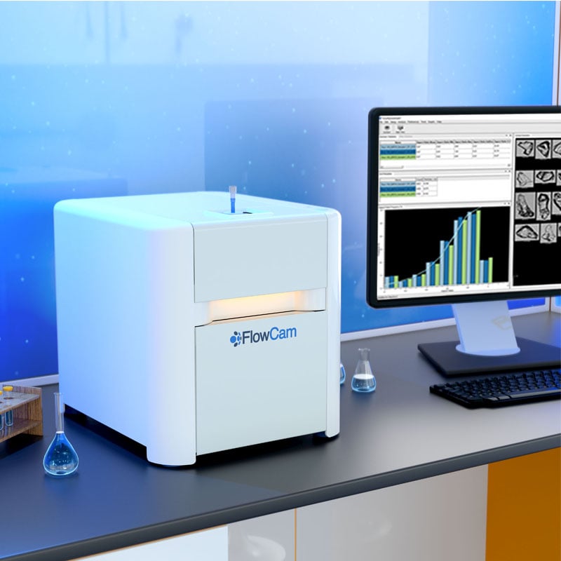
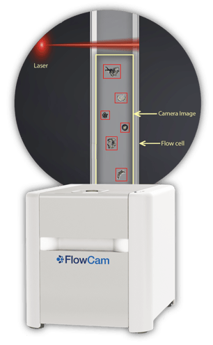
FlowCam, a high-speed FIM instrument, was originally developed to rapidly image and characterize plankton in seawater. Today, it is used across aquatic research, water quality monitoring, biopharmaceutical analysis, and materials characterization to provide statistically robust particle data alongside high-resolution images.
Evaluating FlowCam for Microplastics Detection
A recent study conducted by our FlowCam team investigated whether FlowCam could detect and differentiate common microplastic particles stained with Nile Red, a fluorescent dye frequently used in microplastics research.
The study focused on five common polymer types:
- Polyethylene terephthalate (PET)
- Polystyrene (PS)
- Polypropylene (PP)
- Polyethylene (PE)
- High-density polyethylene (HDPE)
Particles ranged from 5 to 50 μm in size and were analyzed using a FlowCam 8400 instrument equipped with a 532 nm laser and dual fluorescence detection channels.
 Experimental Design
The experimental workflow was deliberately simple and designed to support high-throughput analysis:
- Prepare microplastic particles (5 mg purified polymer from 1 to 50 µm).
- Stain particles using Nile Red in 25% acetone.
- Incubate for 45 min at room temperature.
- Strain to remove excess dye.
- Analyze samples with FlowCam in both AutoImage and Trigger modes.
The use of Trigger Mode allows image acquisition only when a fluorescent particle is detected, helping reduce interference from non-target particles. AutoImage mode captures all particles in a sample.
Nile Red Successfully Enables Fluorescence Detection in Microplastic Particles
One of the first questions addressed by the study was whether unstained particles would generate unwanted fluorescence signals.
The answer was encouraging. Unstained control samples produced no detectable fluorescence in Trigger Mode, indicating that fluorescence signals were associated with Nile Red-stained microplastics rather than background autofluorescence.

We also found that staining efficiency varied by polymer type. Differences in polymer chemistry, polarity, and surface properties appeared to influence how effectively Nile Red bound to each material. Despite these differences, all tested polymer classes produced measurable fluorescent signals that could be detected by the system.
Can FlowCam Differentiate Polymer Types?
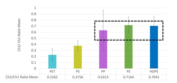
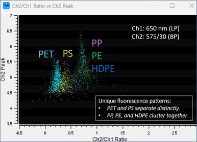
The study demonstrated that fluorescence characteristics provided a useful starting point for discrimination.
PET and polystyrene exhibited distinct fluorescence responses, separating clearly from other polymer groups when fluorescence ratios from the two detection channels were compared. Meanwhile, polypropylene, polyethylene, and HDPE formed a closer cluster.
However, fluorescence alone was not the entire story.
The greatest advantage of flow imaging microscopy is that it provides images and morphology measurements for every detected particle.
In addition to particle concentration, researchers can evaluate size distribution, circularity, symmetry, aspect ratio, fluorescence intensity, and more. And every data point is backed up by an image of the particle that can be used for visual confirmation.
This additional information becomes particularly valuable when attempting to distinguish between different microplastic populations.
Size-Based Differentiation
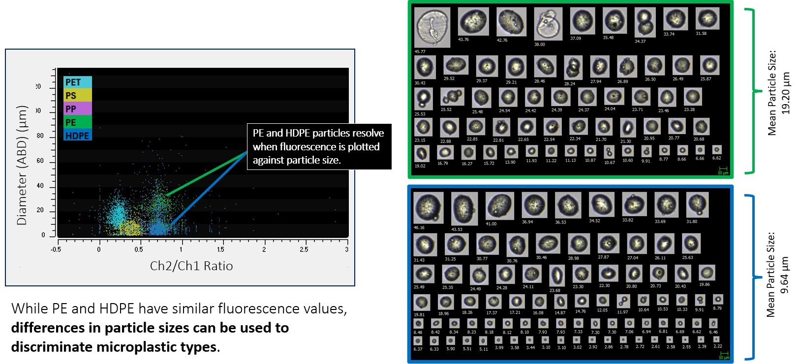
We observed measurable differences in particle size between PE and HDPE populations. By combining fluorescence and size measurements, the two polymer types became easier to distinguish.
Morphology-Based Differentiation
Shape descriptors offered another layer of discrimination.
For example, polypropylene particles displayed lower circularity and symmetry than HDPE particles, creating additional opportunities for classification using image-derived measurements.
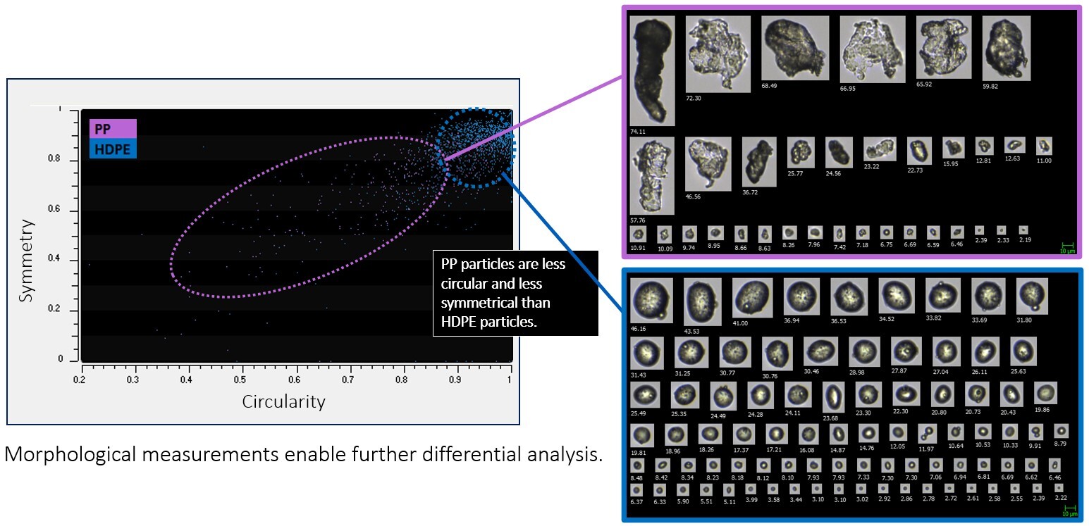
This illustrates one of FlowCam's key strengths: multiple independent measurements can be used together to improve particle characterization.
A Tiered Approach to Microplastics Analysis
FlowCam is not intended to replace FT-IR or Raman spectroscopy for definitive polymer identification. Instead, it can function as an efficient screening technology within a larger analytical workflow.
A practical approach could look like this:
Step 1: Rapid screening with FlowCam to quantify particles and evaluate morphology.
Step 2: Identify samples exhibiting elevated microplastic concentrations.
Step 3: Perform FT-IR or Raman analysis only on samples requiring polymer confirmation.
This strategy enables laboratories to process more samples while reserving resource-intensive spectroscopy methods for the samples that matter most.
Study Conclusions
Based on the findings, our study concluded that FlowCam 8400 equipped with a 532 nm laser is effective for:
- Rapid microplastic screening
- Particle enumeration
- Morphological characterization
- Fluorescence-based discrimination of some Nile Red-stained polymers
The work also demonstrated that combining fluorescence data with particle size and morphology can improve differentiation between microplastic types. While additional validation is needed for complex environmental samples and low-concentration drinking water matrices, the results suggest flow imaging microscopy can play an important role in future microplastics monitoring programs.
What's Next?
Future research will focus on optimizing Nile Red staining procedures and applying this workflow to environmental water samples—some of which is already underway in our application lab! As monitoring requirements continue to evolve, rapid screening tools that provide both particle counts and visual confirmation will become increasingly valuable.
For laboratories seeking a faster way to prioritize microplastic samples for further analysis, flow imaging microscopy offers an attractive balance of speed, sensitivity, and image-based insight.
Wondering if FlowCam will work for your microplastics application? Please reach out—we'd love to collaborate!