


Biopharma
Advanced Subvisible Particle Analysis for Biotherapeutics Development
Biotherapeutics hold great promise, but their complexity makes them especially prone to particle formation. During processing, handling, and storage, these medicines can generate particles that conventional counting methods may overlook. If undetected, such particles can undermine safety, efficacy, and regulatory approval.
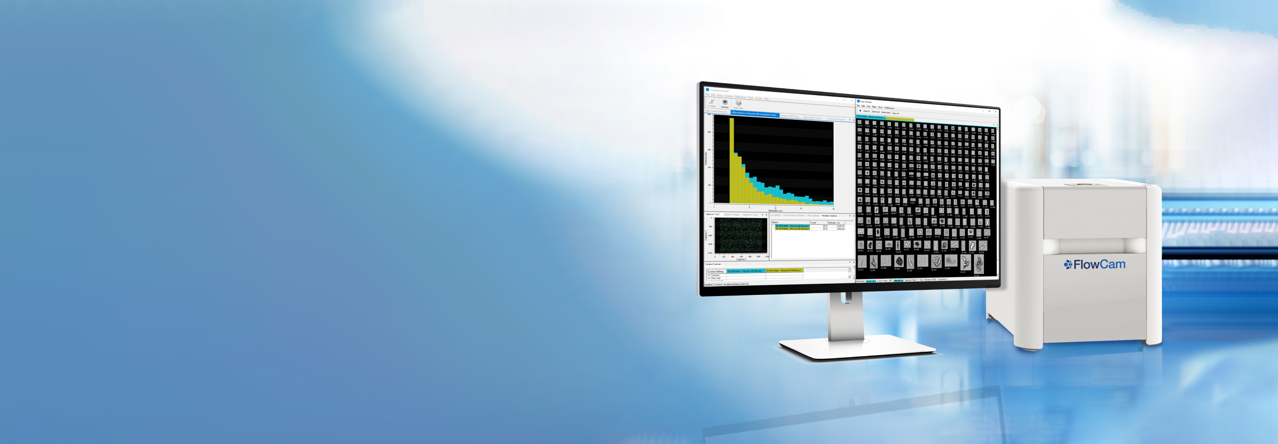
FlowCam delivers clarity in biotherapeutic particle analysis. Combining precise particle concentration measurements with high-resolution imaging, FlowCam reveals both how many particles are present and what they are. This enhanced visibility supports compliance with pharmacopeia guidelines and gives development teams the confidence to optimize formulations and protect patient safety.
Characterize Critical Particles Across All Modalities
Unlike small molecules, biologics are structurally delicate. Manufacturing and storage stresses affect stability, often causing aggregation and generating particles that can trigger immune responses or other adverse effects.
The challenge goes beyond detection—it’s precise characterization. Traditional light obscuration provides counts but lacks the visual detail needed for confident decisions. Without that context, formulation and manufacturing choices may depend on incomplete data, risking delays, failed batches, or worse: safety issues that derail years of development.
“There is great value in seeing images of the particles and of the actual contaminants. It allows us to identify particle type and particle source via morphology, which increases the strength of the finding.”
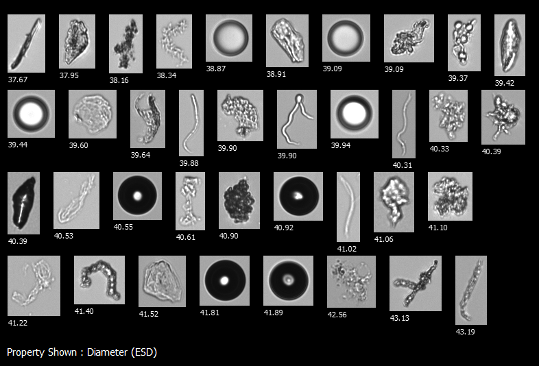
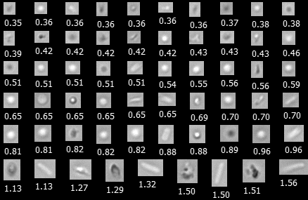
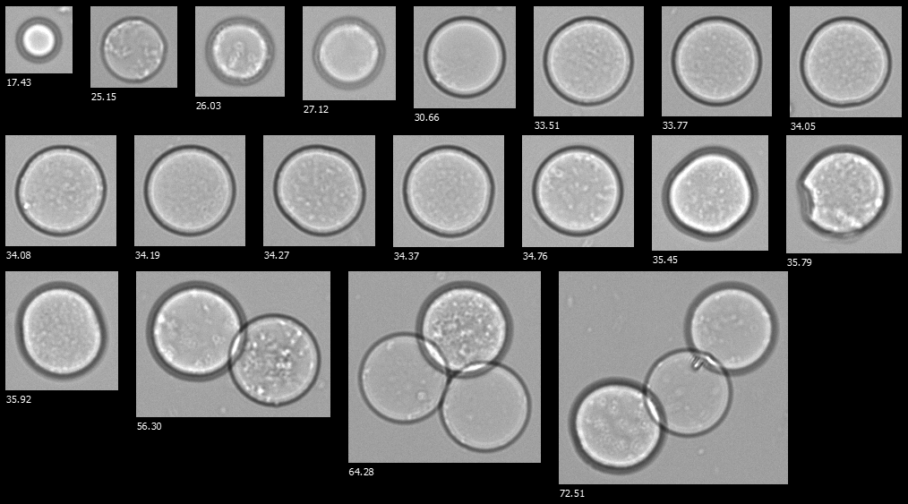
FlowCam images of particles in a biotherapeutic sample, including protein aggregates, silicone oil droplets, fibers, and other contaminants
A FlowCam collage of hydrogel microspheres
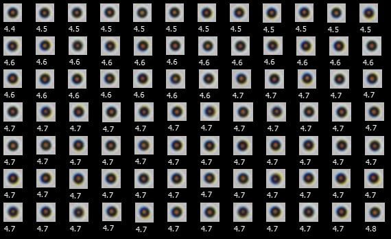
A FlowCam collage of Dynabeads
How FlowCam Strengthens Biotherapeutic Development
FlowCam turns uncertainty into actionable insight by combining quantitative data with morphological confirmation in a single workflow. This dual approach helps biopharma teams see beyond counts to the particle types that truly impact product quality and safety.
With FlowCam, you can:
- Differentiate protein aggregates, silicone oil droplets, and other translucent particles undetectable by light obscuration
- Visualize particle formation to guide formulation development and prevent late-stage surprises
- Monitor particle profiles consistently across development, scale-up, and commercial manufacturing
- Generate comprehensive particle data aligned with USP <788> recommendations using FlowCam LO
- Comply with USP <1788> requirements with FlowCam 8000
- Rapidly identify whether failed batches result from degradation, contamination, or packaging issues
Additional Resources

Interested in learning more?
-
Get in Touch
Tell us about your application and particle characterization needs.
-
Have a Conversation
We're happy to set up a call to discuss your application and answer your questions.
-
Discuss Next Steps
Expand your knowledge with a seminar, demonstration, sample analysis, or obtain a quote.
