Overview
FlowCam Particle Characterization
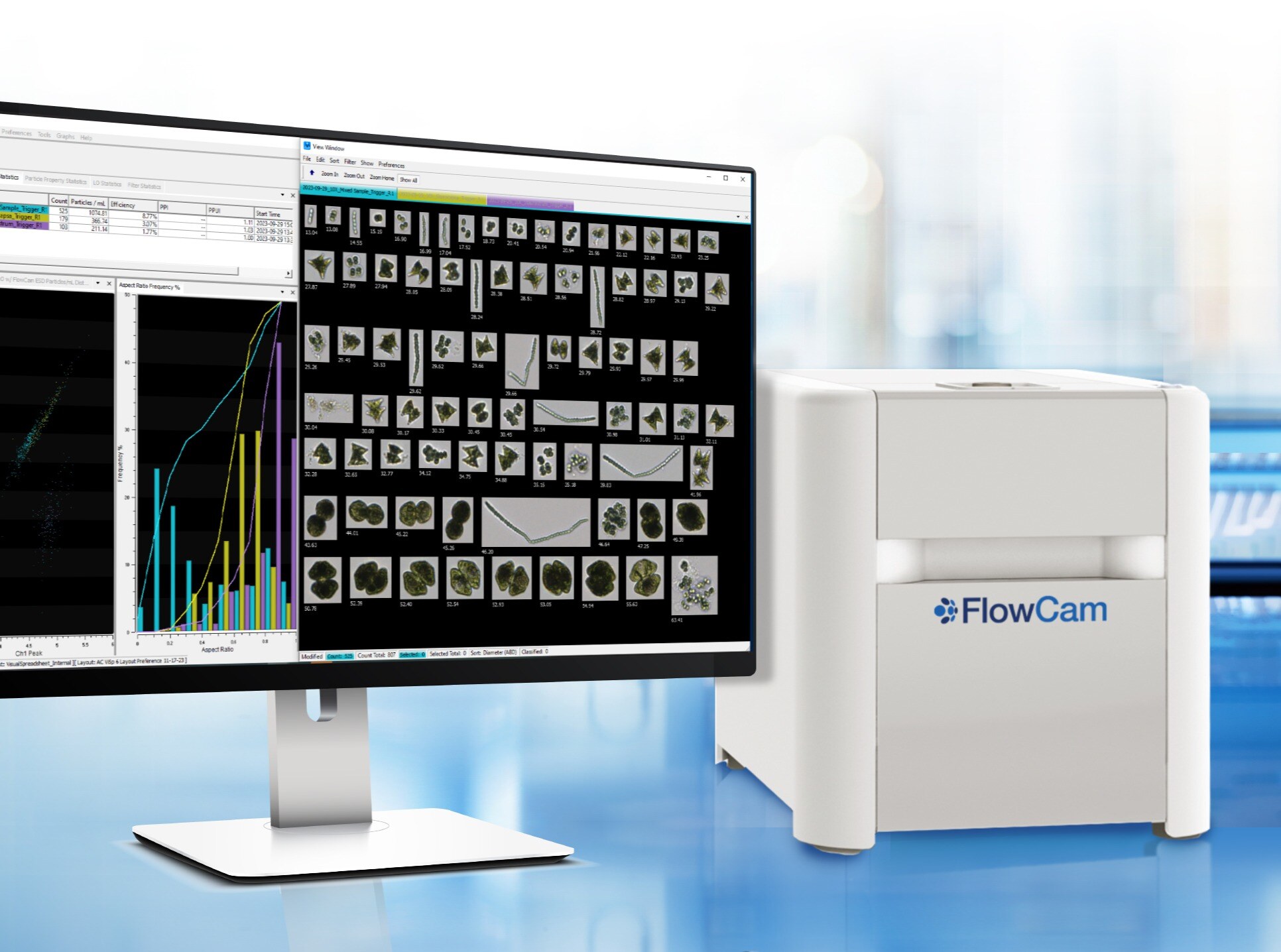
FlowCam 8000 is an image-based particle size and shape analyzer that uses flow imaging microscopy to capture high-resolution images of particles in a fluid medium, combining the benefits of digital imaging, flow cytometry, and microscopy.
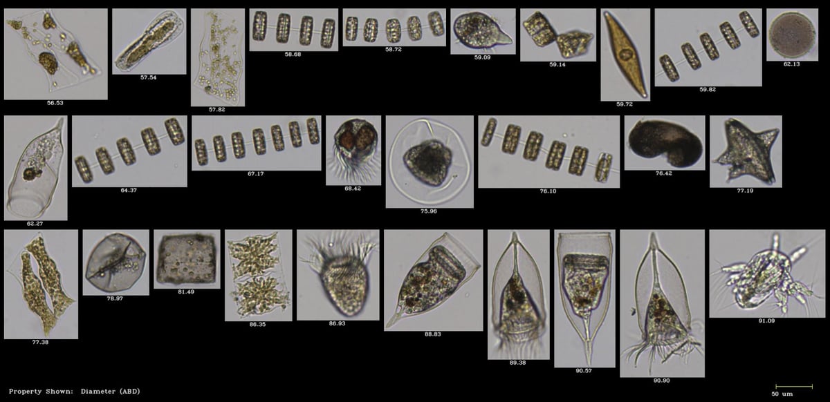
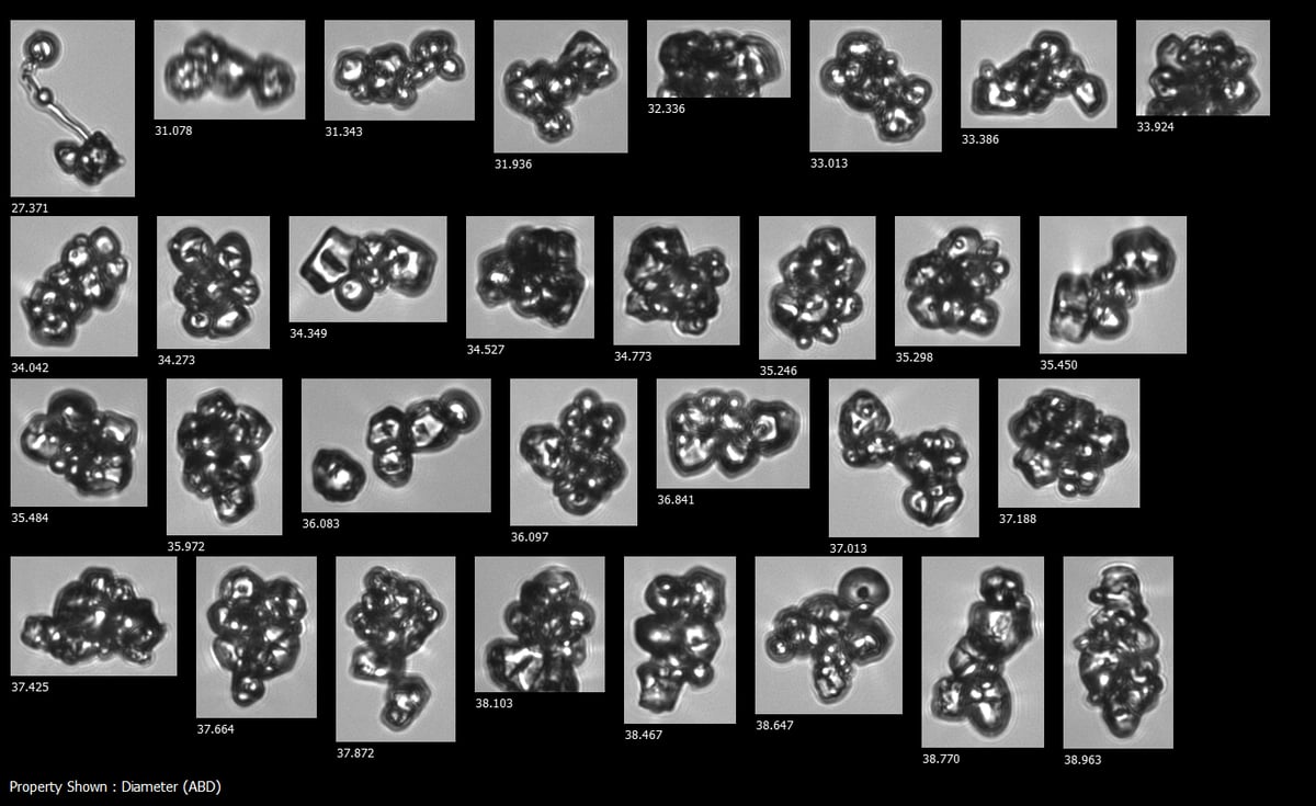
Image-based particle analysis allows for the comprehensive characterization of subvisible particles, API aggregates, and contaminants in biopharmaceuticals, as well as mammalian cells, microplankton, emulsions, and advanced materials.
Benefits
Why use an image-based particle size and shape analyzer?
FlowCam 8000 uses flow imaging microscopy to capture high-resolution images of particles, enabling users to analyze the shape, size, and count of subvisible particles and microorganisms.
- Obtain meaningful results in less than a minute, with as little as 100 μL of sample.
- Distinguish between organisms and other particles by size, shape and color.
- Analyze samples in their native solution, including seawater.
- Differentiate aquatic organisms with fluorescence detection in FlowCam 8400 and FlowCam Cyano.
- Increase productivity with automated liquid handling for up to 384 samples.
Specifications
FlowCam 8000 Series
| Specification |
FlowCam 8100 |
FlowCam 8400 |
FlowCam Cyano |
| Applications |
Biopharma, Aquatic, Materials |
Aquatic |
Aquatic |
| Size Range |
2 µm to 1 mm |
2 µm to 1 mm |
2 µm to 1 mm |
| Magnification |
20X, 10X, 4X, and 2X |
20X, 10X, 4X, and 2X |
20X, 10X, 4X, and 2X |
| Focus System |
Automatic |
Automatic |
Automatic |
| Minimum Sample Volume |
100 µL |
100 µL |
100 µL |
| Flow Rate |
0.05 mL/min to 10 mL/min |
0.05 mL/min to 10 mL/min |
0.05 mL/min to 10 mL/min |
| High-Resolution Camera Type |
Color or monochrome |
Color |
Color |
| Fluorescence excitation |
None |
488 nm or 532 nm with 2-channel fluorescence detection |
633 nm with 2-channel fluorescence detection |
| Automated Liquid Handling Compatible? |
Yes |
Yes |
Yes |
FAQs About Particle Size & Shape Analyzers
What is a particle analyzer?
Particle analyzers are analytical instruments used to examine small objects in a sample. Particle analysis instruments enable scientists to count and characterize particles in a wide range of sample types, including biopharmaceutical formulations, environmental samples, and advanced materials.
What is particle characterization, and why is it important?
Particle characterization refers to the strategies used to distinguish between particle and organism types based on size, shape, color, and other physical properties. This data can help researchers monitor and control unwanted and potentially harmful particle types. Instruments with high-resolution image quality provide reliable measurements of particle size and shape, enabling the distinction between particles with different characteristics.
Why is particle counting important?
The number of particles in a sample must be accurately enumerated to meet regulatory guidelines, ensure human and environmental safety, and quantify ecological changes over time. Instruments that offer accurate and repeatable concentration measurements help ensure that every decision is made with confidence.
What techniques are used for particle size and shape analysis?
Various techniques can be employed to characterize particles, depending on the specific information required and the desired level of automation. Some techniques include:
- Manual microscopy: Uses a brightfield light microscope to view particles in a sample. The magnified images can be inspected to analyze particle shape, color, and morphology.
- Flow cytometry: Particles in a sample are arranged into a single-file line using a sheath fluid and passed through a laser. Each particle scatters light and may fluoresce when passing through the laser, creating light signals that can be recorded to process particle count, size, shape, and structure.
- Light obscuration: Particles in a liquid sample are passed between a light source and a detector. Particles cast shadows on a detector, creating electrical signals that can be used to determine particle concentration and size.
- Electrozone Sensing: Particles suspended in a conductive medium are passed through an aperture located between two electrodes. Each particle generates an electrical signal as it passes through the aperture, which can be analyzed to determine the particle concentration and size.
- Laser diffraction: A liquid sample containing particles passes through a laser. Particles in the sample scatter the laser at different angles depending on their size. The observed scattering angles can be used to determine the particle size distribution present in the sample.
- Flow imaging microscopy: An analytical method to monitor particles. It combines aspects from each of these techniques and offers a way to monitor multiple relevant attributes of a sample’s particle composition with a single measurement.
FlowCam 8000 uses Flow Imaging Microscopy to generate detailed visual and analytical data from particles in fluid samples.
How do I choose the right particle analysis instrument for my research?
The right particle analysis instrument should be able to monitor multiple relevant attributes of the contents of your samples. The size range of the instrument should also align with the sizes of the organisms and other objects you wish to characterize.
What is the value of imaging as it relates to particle size and shape analysis?
Images offer scientists a more comprehensive understanding of the appearance and structural characteristics of particles within a sample beyond the insights provided by basic size and shape measurements alone. Images are also a powerful visualization of the particle types in a sample when processing data, gaining insights, and communicating findings. High-quality particle images are necessary for identifying different plankton taxa, pharmaceutical contaminants, and other complex particle types in samples.


