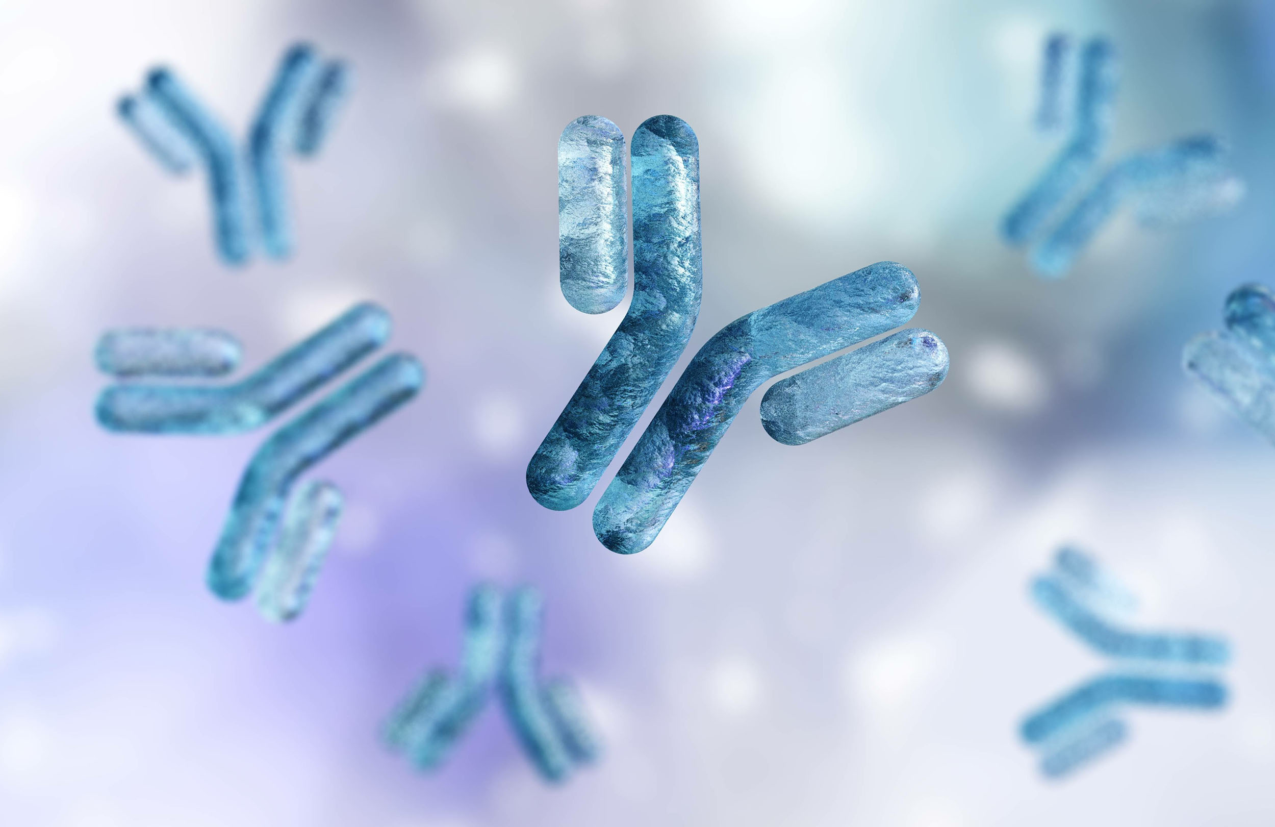
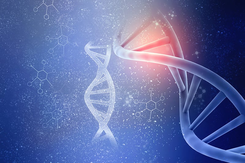
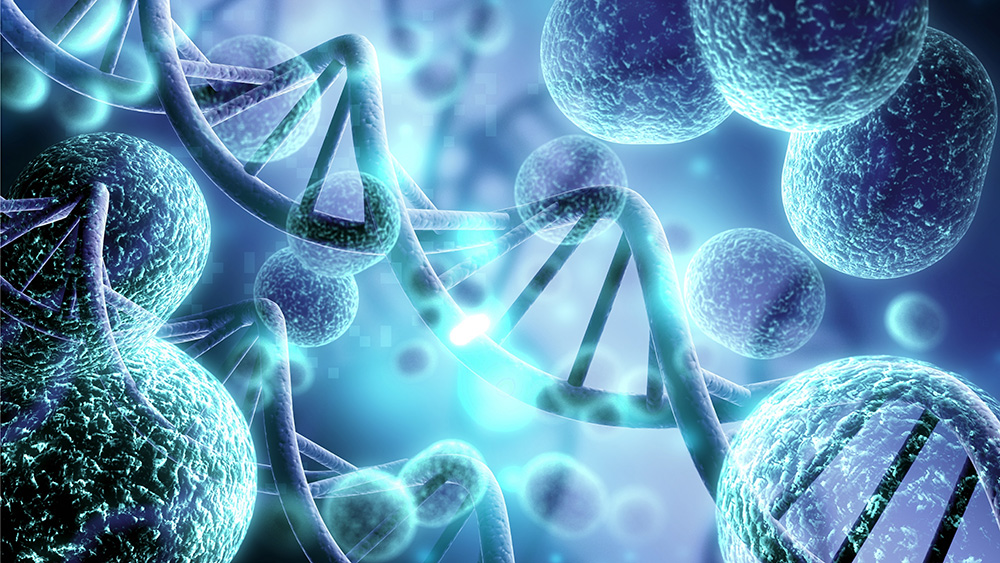
FlowCam is used for quality control and quality assurance in all stages of drug development and manufacturing. Hundreds of biopharmaceutical and pharmaceutical companies worldwide are using FlowCam to improve the quality and safety of their formulations.
FlowCam captures high-resolution digital images of particles in biotherapeutic formulations between 300 nm and 10+ µm in size. FlowCam’s software VisualSpreadsheet analyzes these images to determine the number, concentration, and size distribution of particles in samples. VisualSpreadsheet also offers a variety of image analysis tools to analyze particle morphology including the optional VisualAITM software for identifying images of protein aggregates, silicone oil droplets, and other contaminants in biotherapeutics.
Use real digital images to verify the size, shape, and identity of your particles. Differentiate between air bubbles, silicone oil droplets, protein aggregates, and contaminants, and determine which particles are inherent, intrinsic, and extrinsic.
In order to fully characterize the particles in your parenteral drug formulation, Flow Imaging Microscopy (FIM) is recommended by USP as a high-throughput orthogonal method to non-imaging particle analysis technology like light obscuration and dynamic light scattering.
Flow Imaging Microscopy: How it works >>
“FlowCam paid for itself in just one incident. Suddenly our [parenteral drug] began to show visible particles. So, we FlowCammed a sample from each step of the manufacturing process and saw that one of our filling pumps was shedding metal shards, catalyzing aggregation. We were able to fix the pump and continue with manufacturing. In just that one instance, FlowCam saved us time, money, and enabled manufacturing to continue that same day.”
— Biopharmaceutical FlowCammer, USA, 2018
FlowCam Nano for particles 300 nm - 2 µm
FlowCam LO Obtain Flow Imaging and Light Obscuration data in one
instrument
FlowCam 8000 for particles 2 µm to 100 µm
ALH for FlowCam for automated analysis of up to 384 samples