

Assess Tumoroid Heterogeneity with High-Throughput Flow Imaging Microscopy
Discover a Rapid, Data-Driven Approach to Quantifying 3D Cell Culture Morphology
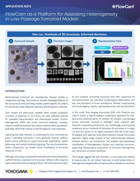
Patient-derived 3D organoid and tumoroid models hold immense promise for personalized medicine and drug discovery. However, characterizing these precious models presents a significant bottleneck. Traditional manual brightfield microscopy is labor-intensive, subjective, and low-throughput, frequently failing to capture the true morphological heterogeneity of low-passage cell clusters.
This application note demonstrates how FlowCam flow imaging microscopy (FIM) transforms quality control and characterization workflows for 3D cell cultures. By combining high-resolution digital imaging with high-throughput flow, FlowCam automatically captures and analyzes thousands of tumoroids directly after their release from extracellular matrices.
In this note, you will explore a proof-of-concept study evaluating four distinct patient-derived tumoroid subtypes. Utilizing FlowCam’s VisualSpreadsheet software, researchers successfully classified complex, heterogeneous cell clusters and sheet-like structures using automated morphological parameters.
Use the form on this page to download the application note and optimize your 3D cell culture workflows.